Palpation is one of the most informative diagnostic methods carried out by a doctor at the stage of the initial examination of the patient. We’ll talk about him today.
Liver anatomy
Before starting to study the question of palpation of the liver, you should decide on the anatomy and functions of the organ. The liver is located immediately below the diaphragm, on the right side at the very top of the abdominal cavity, and only a small part of the organ in the adult is located on the left (in accordance with the middle line). In newborns, the liver occupies a significant part of the abdominal cavity.
Topographically, the liver has two surfaces and two edges. The anteroposterior (upper) surface is adjacent to the surface of the diaphragm and protrudes in accordance with its bend. The lower one is located behind and below, having several impressions from the adjacent organs. The lower and upper surfaces are separated by a lower sharp edge, the other edge (posterior upper), on the contrary, is very obtuse, and therefore it can be attributed to the rear surface of the organ.
There are two lobes in the liver: the greater right and smaller left, separated by a crescent ligament, in the free part of which lies a fibrous dense cord - the so-called circular ligament that stretches from the navel and is nothing more than an overgrown umbilical vein.
The right lobe is divided using furrows into several secondary lobes. In one of these grooves there is a gall bladder and a vena cava (inferior), separated by a piece of liver tissue, which is called the caudate process.
One of the important parts of the organ is the transverse deep groove, which is called the portal of the liver. Through this formation, the large hepatic arteries, portal vein and nerves enter the organ, and the efferent hepatic duct (evacuation of bile into the gallbladder) and lymphatic vessels leave it .
In the right lobe of the organ, a square lobe is distinguished , which is limited by the gates of the liver, the round ligament and the fossa from the gallbladder, and the caudate lobe located between the gates of the liver and the portal vein.
Liver function
- Metabolic (control over the exchange of fluids, trace elements and vitamins, hormones, amino acids, lipids, proteins, carbohydrates).
- Deposited (in the body accumulate BZHU, vitamins, trace elements, hormones).
- Secretory (production of bile).
- Detoxification (carried out thanks to the natural liver filter - hepatic macrophages).
- Excretory (due to the binding of glucuronic and sulfuric acids to toxic substances: indole, tyramine, cattle).
- Homeostatic (involvement of the liver in the control of antigenic and metabolic hemostasis of the body).
Due to morphological and functional features, the liver is often affected in a variety of non-infectious and infectious diseases. That is why, at the first treatment of the patient, palpation of this organ is necessary.
Palpation and percussion of the liver
Before probing the liver, it is recommended that percussion determine its boundaries. This will not only suggest an enlargement of the organ, but also understand where exactly palpation should begin. With percussion, the liver tissue makes a dull (dull) sound, but due to the fact that the lower part of the lung partially covers it, it is possible to determine two boundaries: true and absolute hepatic dullness, but more often they determine only the border (lower and upper) of absolute dullness.
Palpation of the organ (technique)
When probing the liver, certain rules must be observed:
- The position of the subject is lying on his back, while the head is slightly raised, and the legs are barely bent at the knees or straightened. Hands lie on the chest to limit its mobility when inhaling and to relax the muscles of the abdomen.
- The doctor is located on the right, facing the patient.
- The doctor bends the slightly bent right palm flat on the patient’s stomach in the region of the right hypochondrium, three to five centimeters lower than the border of the liver, previously determined percussion. With his left hand, the doctor covers the chest (its lower part on the right), while four fingers must be placed behind, and one (thumb) finger should be placed on the costal arch. This technique will provide immobility of the chest during inspiration and increase the displacement of the diaphragm down.
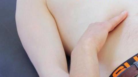
- When the patient exhales, the doctor effortlessly pulls the skin down and, immersing the fingers of his right hand in the abdominal cavity, asks the patient to take a deep breath. At this time, the edge (lower part) of the organ drops, penetrates into the created pocket and slides on the fingers. At the same time, the probing hand should remain motionless. If for some reason the liver could not be palpated, the procedure is repeated, however, the fingers are shifted a few centimeters up. Perform this manipulation, moving higher and higher, until the right hand stumbles on the costal arch, or until the liver edge is palpated.
Features
- Palpation of the liver is usually done along the direct muscle of the abdomen (its outer edge) or the mid-clavicular right line. But if there is such a need, palpation is performed in five lines (from the front axillary on the right to the periosternal on the left).
- In case of accumulation of large volumes of fluid in the abdomen, palpation is difficult. Then they resort to a running ball-jerking probing of the organ. To do this, the second, third and fourth fingers of the right hand perform shock-pushes on the front wall of the abdomen, starting from the bottom and ending with the costal arch until the dense formation is detected - the liver. During a push, the organ first moves deep into the surface, and then it comes back and stumbles onto the fingers (a symptom is called a “floating ice floe”).
Interpretation of results (norm)
What results should palpation of the liver show?
- Normally, in 88% of patients, the lower edge of the organ is located near the costal arch, in accordance with the mid-clavicular line on the right.
- In a healthy person, the edge of the organ is sharp or slightly rounded. It is soft, painless, easily tucks when felt, even.
Evaluation of the obtained data (pathology)
- If the liver is enlarged, on palpation it will be located below the costal arch, which can also indicate its displacement. In support of this or that statement, it is necessary to carry out percussion in order to determine the boundaries of the body.
- If the size of the liver is not changed, but the boundaries of hepatic dullness are shifted down - this is a sign of organ prolapse.
- Displacement of only the lower border indicates an increase in the liver, which occurs with venous stagnation, inflammation in the biliary tract and liver, acute infections (malaria, cholera, typhoid fever, dysentery), cirrhosis (at the initial stage).
- If the lower boundary is shifted up, then a decrease in the size of the organ can be suspected (for example, in the terminal stages of cirrhosis).
- A change in the location of the upper hepatic border (down or up) rarely indicates damage to the organ itself (for example, with echinococcosis or liver cancer). This is often observed due to the high position of the diaphragm during pregnancy, ascites, flatulence, due to the low location of the diaphragm during enteroptosis, pneumothorax, emphysema, as well as in cases of diaphragm moving away from the liver due to gas accumulation.
- Pulmonary infarction, wrinkling of its lower part, pneumonia, right-sided pleurisy can also imitate an upward displacement of the upper border of the organ.
- In some cases, not only palpation of the edge of the liver is available, but also of the whole organ. To do this, the fingers are placed directly under the costal right arch. The doctor, gently pressing, examines the liver with sliding movements, while evaluating its surface (uneven, smooth, even), texture (firm, soft), presence / absence of pain.

- A soft, even, smooth surface and a rounded edge painful on palpation are signs of inflammatory processes in the organ or the manifestation of acute blood stasis due to heart failure.
- A tuberous, uneven, dense edge is observed with echinococcosis and syphilis. A very dense ("wooden") liver is determined when the organ is damaged by cancer cells.
- The dense edge of the liver indicates hepatitis, and in combination with tuberosity - cirrhosis.
- Pain during palpation of the liver can occur due to inflammatory processes or as a result of overstretching of its capsule (with congestive liver).
Palpation of the liver in children
The palpation of the liver of the newborn, as a rule, is carried out at the level of the mid-clavicular, as well as the anterior axillary lines by sliding palpation. In this case, the hand of the examining pediatrician slides off the edge of the liver, due to which it is possible not only to determine the size of the organ, but also to palpate the assessment of its edge. The norm for newborns is the appearance of the hepatic margin from under the costal arch by two (but not more) centimeters. Assessment is carried out on the midclavicular line. The edge of the organ should be painless, smooth, sharp and soft-elastic.
In healthy children up to the age of seven, the edge of the liver, as a rule, protrudes from under the costal right arch and is accessible for palpation. For healthy children under three years of age, the definition of the edge of the liver 2 or 3 centimeters below the right hypochondrium is considered normal. After seven years, the borders of the liver correspond to those in adults.
The study of the liver Kurlov method
To confirm the diagnosis of a particular pathology, which leads to a distortion of the size of the organ, it is necessary to carry out palpation of the liver according to Kurlov. To do this, using tapping (percussion) determine the upper border, and then palpation (or percussion) - lower. Moreover, in accordance with the oblique stroke of the lower edge of its boundary, as well as the distance between the upper and lower boundaries, are determined by three points.
The first corresponds to the midclavicular line, the second to the midclavicular, and the third to the costal left arch. In the room, the sizes should be 9, 8, 7 cm, respectively.