Vitreoretinal surgery is a complex progressive area of ophthalmic surgery, which involves an integrated approach for the treatment of diseases of the posterior region of the eyes, that is, we are talking about the retina and vitreous. As a result, experienced surgeons solve not just difficult clinical tasks, but correct pathological conditions that were previously considered hopeless.
Vitreoretinal eye surgery - what is it?
The medical term "vitrectomy" comes from two concepts - "vitreum", which means the vitreous body, and "ectomy", translated as removal. Thus, vitrectomy involves the surgical removal of the vitreous body of the left or right eye. Such an operation is performed in ophthalmology departments.
Vitreoretinal eye surgery typically involves vitrectomy. Vitrectomy is a minimally invasive high-tech seamless operation aimed at removing the vitreous body that fills the cavity of the eye. Surgical function is performed through micro-punctures. This operation allows you to effectively treat various diseases of the vitreous body and retina.
The purpose of its implementation is the prevention of vision loss along with the restoration of the anatomical ocular structure.
Let us consider in more detail the history of vitreoretinal surgery.
Von Graefe and Deutschmann were the first to offer retinal detachment treatment. They dissected vitreal membranes with a knife. The Nedden method was also used - a clouded vitreous body was removed by suctioning it from the eye with a special needle and syringe.
In 1971, R. Machemer was the first to perform a vitrectomy through the flat part of the ciliary body with controlled intraocular pressure. For this, a vitreot with a diameter of 1.5 mm was developed. Due to the desire for improvement, the development of R. Kloti (1973) of the electric guillotine type vitractor appeared.
Then, the procedure for the operation of segmental filling without drainage was allocated to the minimal surgery of retinal detachment. In 1974, Heintz was the first to use the 3-port vitrectomy method.
Indications for
Vitreoretinal surgery is used in a number of the following situations:
- With retinal detachment.
- Against the background of the tension of the retina by the vitreous body, which leads to its detachment, especially when it can affect the central zone, which is responsible for objective vision.
- Against the background of retinal breaks.
- With hemophthalmia (vitreous hemorrhage). This operation allows you to reduce the loss of vision, which is caused by the process of hemorrhage in the vitreous body, when the bleeding is plentiful or when the blood cannot be resorbed for several months.
- Against the background of injuries and surgical operations affecting the posterior ocular segment.
- In the presence of a foreign body inside the vitreous.
- Against the background of significant opacification of the vitreous body.
- In the presence of severe proliferative retinopathy, causing the formation of fibrous tissue on the retina.
- Overgrowth of new blood vessels (neovascularization), despite repeated laser treatment.
Preparation for surgery
Preparation for vitreoretinal surgery for retinal detachment, as a rule, involves a thorough examination of the organ of vision, as well as an assessment of the general condition with the presence of concomitant pathologies in the patient. The diagnostic algorithm directly depends on the pathological condition in relation to which the operation is planned. Preparation may include:
- Perform inspection with a slit lamp.
- Ophthalmoscopy with dilated pupil.
- Carrying out optical coherence tomography.
- Perform fluorescence angiography.
- Conducting ultrasound examination of the retina.
Advanced diagnostics are necessary when planning the involvement of the anterior segments of the eyes, cornea or lens during an operation. In the event that there has been one or another traumatic damage to the organs of vision, then computer or magnetic resonance imaging may be required. These imaging techniques are needed to assess the extent of the injury.
After diagnosing a disease that requires the use of vitreoretinal surgery, the doctor will explain to the patient information about the indications, risks and alternatives. After this, the patient signs an informed consent for surgery.
Eight hours before surgery, it is recommended to stop drinking and eating. This minimizes the risk of aspiration of the contents of the stomach during anesthesia. In the event that a person uses some drugs constantly, then their preoperative use must be agreed in advance with a specialist. Medications such as injectable insulin along with anticoagulants or antiarrhythmic drugs should be discussed in detail with the surgeon or anesthetist.
Types of Vitrectomy
Depending on the scope of the intervention, vitreoretinal surgery can have several varieties:
- The total type of operation in which the entire vitreous volume is removed to the patient.
- Subtotal version of surgical intervention, against the background of which a particular segment is removed. For example, in the presence of vitreoretinal traction in a patient, the posterior vitreous segments are removed.
Vitreoretinal Surgery Technology
During vitrectomy, the surgeon introduces the finest instruments into the eye, cuts off the micro-pieces of the vitreous body, and it is aspirated. After the vitreous removal procedure, the surgeon proceeds to the treatment of the retina with a laser (i.e., laser endocoagulation is performed). As part of the technology, fibrous or scar tissue is excised or removed, the area in which the retina is exfoliated is straightened, traction is removed and the hole in the retina or macula is removed.
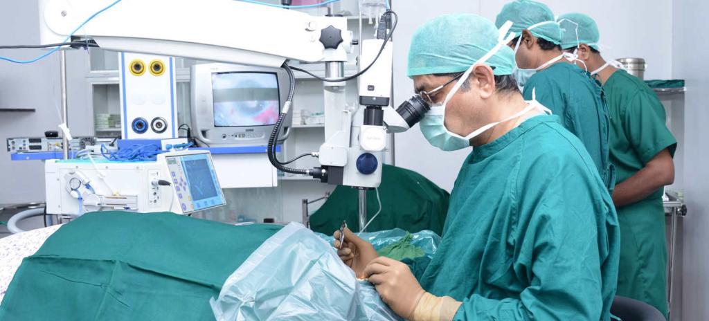
A photo of vitreoretinal surgery is presented.
At the end of the operation, in order to replace the vitreous body, and at the same time restore normal pressure in the eye, a special balanced salt solution is introduced along with silicone oil or gas.
A vitrectomy is always performed by a surgeon who has special training in the treatment of retinal problems. It is worth noting that most modern ophthalmologists are highly professional specialists in the field of microsurgical operations.
What to expect after such an operation?
Vitreoretinal surgery may be performed on an outpatient basis, but hospitalization may be required in some situations. The operation, as a rule, lasts from one to three hours, and it is performed under local or general anesthesia, depending on the recommendations of the anesthesiologist and surgeon.
After the operation under domestic conditions, for a short time, a person will have to adhere to a certain position of the head. The surgeon will tell you which one, depending on the disease and on what exactly the patient has replaced the vitreous body.
Treatment effectiveness
Vitrectomy can significantly improve visual acuity among patients suffering from vitreous hemorrhage, which cannot be resolved. The operation helps to reduce the risks of serious complications of chronic bleeding into the vitreous body and the proliferation of blood vessels. Vitrectomy is sometimes the only method for eliminating proliferative vitreoretinopathy and retinal detachment.
The operation helps to restore vision, which is lost in the process of traction detachment. It also prevents further delamination. But the result, as a rule, will be better when the detachment does not affect the center of the retina.
The risks
Vitrectomy can cause an increase in pressure inside the eyes, especially in people with glaucoma. There are several other complications that are associated with vitrectomy. These include:
- Prolonged bleeding.
- Relapse of retinal detachment along with corneal edema.
- Infections inside the eyes, that is, the development of endophthalmitis.
Subtleties
Vitreoretinal surgery is performed exclusively on modern surgical equipment. All methods are minimally invasive, less traumatic, and for the most part they are all seamless and painless.
Monitoring of the patient's general well-being during the operation is carried out by experienced anesthetists. All risk is minimized through the use of expensive disposable supplies.
In what cases are methods of such intervention used today?
This type of surgery is applicable in the following cases:
- In the event that the vitreous volume is lost as a result of a traumatic effect on the eye or as a result of a previous intervention.
- When the vitreous body of the patient is cloudy.
- If the patient is diagnosed with maculopathy along with retinopathy with diabetes.
- In the event that a person has retinal detachment. Only a doctor can make the right and informed decision about vitreoretinal surgery.
- In the event that, against the background of a general illness of the body, the patient has a repeated vitreous hemorrhage.
- When there is a macular rupture along with macular degeneration.
- If you want to correct a complication after a previously performed operation aimed at removing cataracts.
- In the presence of injuries of the eyeball.
In case of eyeball contusion or against the background of a penetrating wound, the question of the preservation of visual functions arises. When conducting timely surgical intervention using the methods of vitreoretinal surgery, in many situations it is possible to maintain high visual function.
Consequences and features of vitreoretinal operations
It is important to understand that the result of vitreoretinal surgery does not appear immediately after the surgery. Naturally, the patient will immediately feel the improvement in the quality of his vision (with the exception of situations of gas injection, against which first you need to wait for its complete evaporation). True, it will be possible to fully evaluate the effect of the operation after two or three months, and sometimes vision can improve within only one year after surgery.
Then we learn what patients who have undergone such treatment talk about vitreoretinal surgery.
Reviews
People who have gone through such an intervention say that in the first few hours immediately after the operation, patients must be in bed. For the period of formation of adhesions, it is required to completely exclude any physical activity, and in addition, refrain from watching TV, playing a computer, reading, and the like.
Also, former patients share that daily washing after surgery should be carried out with the head tilted back, this completely eliminates the risk of water entering the operated eye. And if water does get in, then it is advised to rinse your eyes with an aqueous solution of "Furacilin" or "Levomycetin." To protect from bright light and dust, it is advised to use a blindfold.
In the reviews, people say that, in general, the rehabilitation period is well tolerated. Postoperative drug treatment, as a rule, consists of antibiotics that help prevent a particular infectious complication. In addition, former patients in reviews of vitreoretinal surgery write that doctors prescribed them anti-inflammatory drugs designed to reduce intraocular pressure and swelling.
According to the reviews of former patients, visual acuity is finally established two or three months after the operation, but for the period of early rehabilitation, temporary glasses are needed along with lenses in order to eliminate eye strain.
Guaranteed Result
The experience of modern surgeons in combination with accurate diagnostic equipment and the best technical equipment of current ophthalmological clinics allows us to predict, and at the same time achieve a high result that exceeds even patient expectations.
Thus, many modern centers of ophthalmologic therapy give their patients a guarantee of obtaining the expected result of the operation. As doctors write in their comments, the safety and comfort of patients is ensured at all stages, whether it is the first examination of the patient or postoperative support.
Vitreoretinal surgery in Belgorod is very popular. You can contact, for example, the clinic "Euromed", a medical center "Harmony of Health" or other institutions.
Postoperative period, or when patients recover vision?
As the surgeons assure, the patients will not have eyes hurt either during or after the surgery. Absolutely all procedures are usually performed on an outpatient basis using local anesthesia, and vision begins to recover in patients immediately on the day of surgery.
There are limitations with recommendations for the period after vitreoretinal surgery in the clinic and in the home, which are explained individually for each patient, which largely depends on the complexity of the intervention.
As a rule, in all specialized ophthalmological centers, patients are given the necessary medications after surgery, and any consultations along with examinations during the rehabilitation month are included in the cost of treatment, if we are talking about paid clinics.
We have considered that this is vitreoretinal eye surgery.