During evolution, the skeleton of the human limbs has undergone significant changes. So, the legs, performing tasks of movement and support, ensure the preservation of the body in an upright position, and the hands become tools of labor. Next, we consider in more detail the skeleton of the upper limb: the structure and tasks that it performs.
General information
The skeleton of the upper extremities of a person acquired significant mobility during phylogenesis. Due to the presence of the clavicle, which provides the connection of the bones of the arms and trunk, people can perform quite extensive movements. In addition, the elements included in the skeleton of the free upper limb have a movable joint with each other. This is especially true for the area of the hand and forearm. The functions of the skeleton of the upper limbs are quite extensive. Hands are adapted to complex types of work. Due to the presence of a large number of bones and joints, the fingers can perform different tasks: from writing to assembling any mechanisms. The leg, acting as an organ for moving and supporting the body in space, includes more massive and thick bones. Their mobility in relation to each other is less significant. The skeleton of the upper and lower extremities is made according to the general plan. It includes two parts.
Parts of the skeleton of the upper limb: bones of the belt
This part includes:
- Shovel. It is presented in the form of a triangular flat bone. Three edges stand out in the scapula. In particular, the lateral, medial and upper parts are present. Also, three angles are distinguished in the scapula. One is lower, the second is medial, and the third is lateral, respectively. The shoulder blade is adjacent to the back of the chest. It is located in the zone between II-VII ribs. In the segment there is an articular cavity. It serves to connect with the clavicle. A protrusion is visible from the back of the scapula. It is located transversely. This protrusion is the spine of the scapula separating the infraspinatus and supraspinatus fossa.
- Collarbone. It is presented in the form of an S-shaped tubular bone. It distinguishes between two ends - the humeral (acromial) and the sternum, and the body. The clavicle is the only element due to which the skeleton of the upper extremities of a person is connected with the bone structure of the body. The sternal end of the segment is thickened. It articulates with the handle of the sternum. The flat shoulder end connects to the shoulder blade.
The second part of
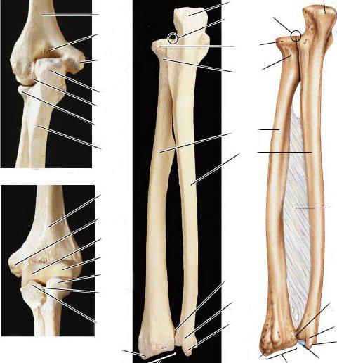
In it, the skeleton of the upper limb consists of a brush, forearm, and shoulder element. The last segment is represented by a single bone - the humerus. The brush includes the finger phalanges, metacarpus and wrist. Two elements are distinguished in the forearm. It is represented by the ulnar and radial bones.
Brachial bone
It is presented in the form of a tubular long element. In the bone, the diaphysis (body) and 2 epiphyses (end) are distinguished: upper and lower. The first is a rounded articular head. It serves to connect with the scapula. The upper end is separated from the body using the anatomical neck. Below it, on the outside, there are tubercles (elevations) - small and large. They are separated by a furrow. The narrowed part in the body closer to the head is called the "surgical neck". On the surface of the bone there is also tuberosity. It acts as a site for joining the deltoid muscle. The lower pineal gland has an extension and passes into the condyle. It serves to connect in the joint with the radius and ulna.
Forearm
In this part, the skeleton of the upper limb includes two elements:
- The ulna. It runs along the inner surface of the little finger (V finger). The upper end of the element is more massive. Two processes are distinguished in it: behind the ulnar and in front - the coronoid. They are separated by a block-shaped notch to connect to the shoulder bone. The outer (lateral) side of the coronoid process contains a radial notch. It forms a joint with a circle. At the lower end, the ulnar bone forms the head. It distinguishes the articular surface in the form of a circle for connection with the ulnar notch in the beam element. The styloid process runs along the inner (medial) side.
- Radial bone. It is presented as a long tubular element. The radius passes along the outer surface from the side of the thumb (first) finger. Its upper end is formed by a cylinder head. There is an articular fossa and a circle on it. The upper ends in the radius and ulna participate in the formation of the joint. The lower section has an elbow notch, an awl-shaped lateral process. There is also a carpal joint surface. The lower sides of the radius and ulna form the wrist joint with the upper row of the carpal bones.
Brush
The skeleton of the upper limb in this area is represented by the bones of the wrist, metacarpus and fingers. The first zone consists of two rows of spongy short bones (four in each). The bones in the wrist are articulated. The upper side of the first row has connections with the radius, through the articular surface. The lower part of the second joins the base of the metacarpal elements. The metacarpus is represented by five tubular short bones. Count start from the thumb. Each metacarpal bone has a head, base, and body. The first element articulates with the upper phalanx in the corresponding finger. The phalanges are tubular short bone elements. They have a head, base and body. The first two elements distinguish articular surfaces. In the upper phalanges, this segment has an articulation with the head in the corresponding metacarpal bone, in the lower and middle ones - with the phalange located above (proximal). There are two tubular bones in the thumb, and three in the rest.
Age-specific developmental features: bone belt
All elements included in the skeleton of the upper limb, except the clavicle, pass through the connective tissue stage, cartilage, and bone.
- Shovel. Its primary site of ossification is laid in the second month of fetal development. From this point, the awn and body of the segment develop. By the end of the 1st year of life, an independent ossification site is laid in the coracoid process, and by the age of 15-18 it forms in the acromion. In the 15-19 year, the coracoid process merges with the scapula.
- Collarbone. Its ossification occurs quite early. This section of the skeleton goes through one - connective tissue - stage. At the 6-7th week. an ossification point appears inside the womb. It is located in the center of the connective tissue primordium. From this site, the formation of the acromial end and body of the clavicle, which in newborns almost entirely consists of bone tissue, takes place. Cartilage forms in the sternum. The core of ossification in it appears only by the age of 16-18, which fuses with the bone body in 20-25 years.
- Shoulder. In the proximal epiphysis, the formation of secondary ossification points occurs: in the large and small tubercle by the 1st-5th, and in the head, usually at 1 year of age. At the age of 3-7 liters. they merge, and 13-25 years - joining the diaphysis. In the distal pineal gland, the site of ossification forms up to 5 years of life, the lateral epicondyle - up to 4-6, the medial - up to 4-11. By 13-21, all parts are fused with the diaphysis.
middle part
- Elbow bone. At 7-14 years in the proximal pineal gland an ossification point is laid. From it begins the ulnar process, in which there is a block-shaped notch. By 3-14 years, sites of ossification in the distal pineal gland are formed. Overgrowing, bone tissue forms the styloid process and head. Fusion with the body of the proximal pineal gland occurs on the 13-20th year, distal - 15-25th.
- Radius. By 2.5-10 years, the site of ossification in the proximal pineal gland is laid. Fusion with the diaphysis occurs by 13-25 years.
The development of brush elements
- Wrist. After birth, ossification of cartilage begins. By the 1st-2nd year of life, the ossification point forms in the hook-shaped and capitate bones, on the 3rd — in the trihedral, the 4th — on the lunar, the 5th — on the scaphoid, the 6–7th — on the trapezoid, on the 8th — in pea-shaped.
- Metacarpus. The laying of the bones that form this part occurs earlier than in the wrist. The ossification zones in the diaphysis are laid by the 9-10th week of embryonic development, with the exception of the first bone. In it, the site forms by 10-11 weeks. In the pineal glands, ossification zones appear between 10 months. and 7 years. By the age of 15-25, the diaphysis and the head of the metacarpal bone fuse.
- Phalanxes. The ossification points of the bodies of the distal elements appear toward the middle of the second month of embryonic development, the proximal ones at the beginning, and the middle at the end of the third month. In the phalanx bases, sites appear between 5 months. and 7 years. The increment to the body occurs in the year 14-21.
The skeleton of the upper extremities has a complex structure in which each element plays a role.