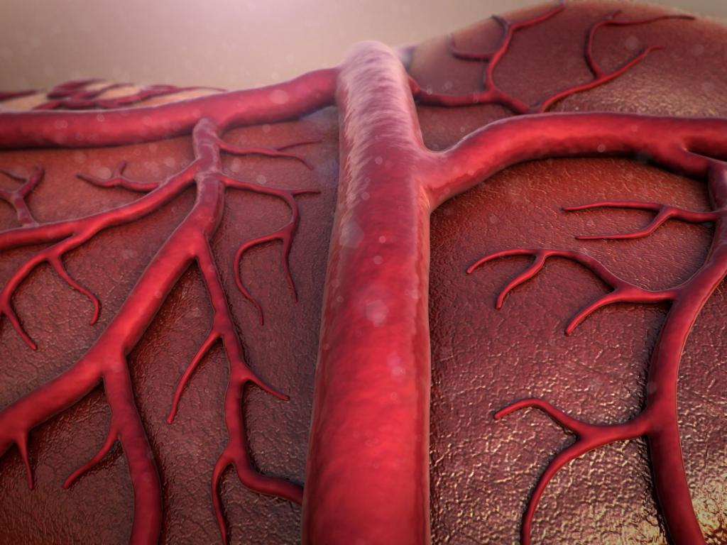
The human body consists of biological tissues penetrated by a mass of blood vessels. They are responsible for the nutrition of cells and the removal of metabolites, supporting their vital functions. Arteries are a type of blood vessel through which blood is delivered directly to the capillary bed. From them through the interstitial fluid, dissolved cells get all the cells of the body.
Morphology
An artery is an anatomical structure in the form of an elastic tube with a wall and a lumen. It passes in the body cavities or connective tissue veins of the parenchymal organs, where it constantly gives up small branches to nourish the surrounding tissues. An artery is a vessel that constantly conducts a pulse wave.
In large vessels, its distribution is achieved mainly due to the elastic qualities of the wall, and in small vessels due to muscle contraction. Like the heart, arterial vessels are constantly in good shape and experience periods of extension and contraction. The muscular wall also alternates with periods of contraction with relaxation.
Histological structure
Any artery is a formation with a multilayer wall, which consists of interwoven elastic fibers and muscle cells embedded between them. This is the structure of the middle wall of the vessel, which is internally covered with a connective tissue membrane. It is based on the endothelial layer facing the inside of the vessel. It is a single-layer simplest epithelium, the cells of which are snug against their edges in order to prevent platelet cells from connecting to the connective tissue membrane. The latter contains platelet adhesion receptors, which is the basis of the thrombus formation mechanism when the endothelial layer is damaged.
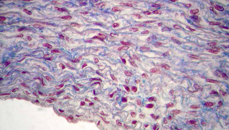
Outside the middle membrane, represented by smooth muscle cells woven into the elastic network, is another layer of connective tissue. It serves to ensure the mechanical strength of the artery. What is it from the point of view of histology? This sheath is a robust network of collagen fibers with integrated single cells. It is connected to the looser adventitia membrane connecting the artery to the stromal tissue of the parenchymal organs.
Arterial tone regulation
All arterial vessels of the body have their own blood circulation, since only the endothelium can be fed from the blood in their lumen. These vessels and nerves pass in the outer connective tissue membrane and supply the middle layer - muscle cells. The smallest nerves of the autonomic system also go to them. They transmit sympathetic impulses that accelerate the conduct of the pulse wave while increasing the heart rate.
In addition, an artery is a hormone-dependent structure that expands or contracts depending on the presence of humoral factors: adrenaline, dopamine, norepinephrine. Through them, the body regulates the tone of the entire vascular system. The main goal is to quickly increase blood flow in the muscles, expanding the blood vessels of the periphery in case of suprathreshold stress. This is an evolutionary mechanism for saving the body’s life by fleeing danger.
The main arteries of the body
The largest artery that can withstand maximum pressure is the aorta - the main vessel, from which the regional branches depart. The aorta originates in the left efferent tract of the corresponding ventricle. In the right efferent tract of the heart, the pulmonary artery originates. This system demonstrates the separation of blood circulation circles: the aorta carries blood into the large circle, and the pulmonary trunk into the small circle. Both of these vessels divert blood from the heart, and veins deliver it to it, where the circulatory system crosses.
Among the most important arteries of the body, renal, carotid, subclavian, mesenteric, iliac arteries and vessels of the limbs should be distinguished. Separately, though not the largest, but extremely important for the body, coronary arteries. What does this mean and why are they special? Firstly, they nourish the heart and form two mutually perpendicular circles of blood circulation of this organ. Secondly, they are special for the reason that these are the only arterial vessels, the filling of which occurs in the ventricular diastole before the development of the pulse wave of the ascending aorta.