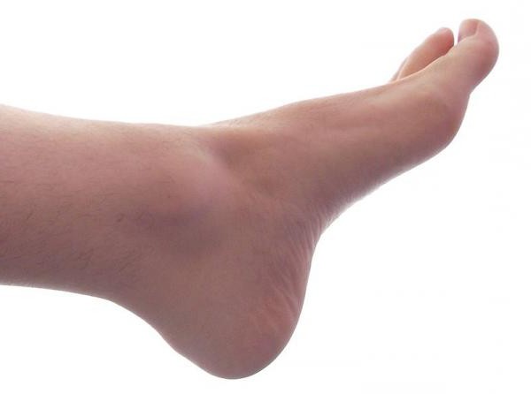
The human foot is that part of the human body that maximally distinguishes bipedal people from primates. Every day she experiences a huge load, so the vast majority of people in one way or another have problems associated with it. A person's foot is constantly influenced by several factors: physical activity, general health, type of profession, shoe pressure. All of them can lead to diseases of this organ.
To understand how you can reduce the negative impact of various factors that affect the health of the lower extremities, you should study what a person’s foot consists of. This organ includes joints and bones; tendons and ligaments; nerves; muscle blood vessels. The human skeleton consists of 26 bones, grouped into 3 sections: proximal (tarsus; talus; scaphoid; calcaneal; cuboid; intermediate, medial and lateral sphenoid bones), metatarsus (consists of five short bones located between the phalanges and tarsus), fingers (14 bones making up segments (phalanges)). It is noteworthy that the thumb has 2 phalanges, and all the others - 3.
The human
foot anatomy is due to our ability to walk on two legs. What is this organ? Its bone basis is the talus, which is tightly connected with the tibia. They form the
ankle joint. The main load of body weight falls on the calcaneus and
metatarsal bones. Five metatarsal bones are connected to the
phalanges of the fingers. The skeleton of the foot is connected by ligaments responsible for the integrity of the joints. An important role is also played by tendons, which combine muscles with bones. Collagen fibers give strength and elasticity to these tissues, which are intertwined in the form of a rope. The most important tendon of the foot is the so-called "Achilles", which is a continuation of the calf muscle. It is attached to the
calcaneus. It is it that allows a person to rise on toes and bend their feet. The tendon extending from the posterior tibial muscle provides turns inward, supports the arches of the foot. Small ligaments connect the bones. Some of them form a capsule surrounding the articular parts of the bones. It is filled with joint fluid.
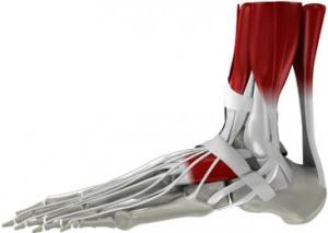
The human foot includes many muscles: the soles, the rear, the intertarsal. Among them are flexion and extensor. In their thickness are nerves, the main of which is the tibial. Going down from under the inner ankle, he appears on the foot. This nerve provides movement of a large number of muscles of the foot, gives it sensitivity. Well, two arteries are responsible for the blood supply to this organ: the posterior and anterior tibial. One of them passes in the sole and is divided into two branches there. The second (front) passes in front of the foot, forming an arc. Venous outflow is carried out using two superficial (large and small saphenous) and two deep veins (posterior and anterior tibial).
In the human foot there are special "arches". It is built like an elastic arch ending in the toes and the heel. The bones of the foot are two arches - transverse and longitudinal. In a healthy person, the load on the lower limb is distributed evenly and in accordance with the phases of a step or run. The human foot rests on the floor behind (calcaneal tuber) and front (metatarsal bones), which provides it with good spring properties.