The uterus is the reproductive unpaired internal organ of the female. It consists of plexus of smooth muscle fibers. The uterus is located in the middle of the pelvis. It is very mobile, therefore, relative to other organs, it can be in different positions. Together with the ovaries, it makes up the reproductive system of the female body.
General structure of the uterus
This internal muscular organ of the reproductive system has a pear-shaped shape, which is flattened in front and behind. In the upper part of the uterus, the sides have branches - the fallopian tubes that pass into the ovaries. Behind the rectum, and in front of the bladder.
The uterine anatomy is as follows. The muscular organ consists of several parts:
- The bottom is the upper part, which has a convex shape and is located above the line of discharge of the fallopian tubes.
- The body into which the bottom smoothly passes. It has a conical shape. Narrows down and forms the isthmus. This is the cavity leading to the cervix.
- Cervix - consists of the isthmus, cervical canal and vaginal part.
The size and weight of the uterus are individual. The average values of its weight in girls and nulliparous women reach 40-50 g.
The anatomy of the cervix, which is a barrier between the internal cavity and the external environment, is designed so that it protrudes into the front of the vaginal fornix. At the same time, its posterior arch remains deep, and the anterior one - on the contrary.
Where is the uterus?
The organ is located in the pelvis between the rectum and the bladder. The uterus is a very mobile organ, which, in addition, has individual characteristics and pathologies of the form. Its location is significantly affected by the condition and size of neighboring organs. The normal anatomy of the uterus in the characteristic of the space occupied in the small pelvis is such that its longitudinal axis should be oriented along the axis of the pelvis. Its bottom is tilted forward. When the bladder is full, it moves back a little, when empty, it returns to its original position.
The peritoneum covers most of the uterus, except for the lower part of the cervix, forming a deep pocket. It extends from the bottom, goes to the front and reaches the neck. The back part reaches the vaginal wall and then goes to the front wall of the rectum. This place is called the Douglas space (indentation).
Uterine anatomy: photo and wall structure
The organ is three-layered. It consists of: perimetry, myometrium and endometrium. The surface of the uterine wall is covered by the serous membrane of the peritoneum - the initial layer. At the next - average level - the tissues thicken and have a more complex structure. Plexuses of smooth muscle fibers and elastic connective structures form bundles that divide the myometrium into three inner layers: inner and outer skew longitudinal, circular. The latter is also called the average circular. He received this name in connection with the structure. The most obvious is that it is the middle layer of the myometrium. The term "circular" is justified by a rich system of lymphatic and blood vessels, the number of which increases significantly as you approach the cervix.
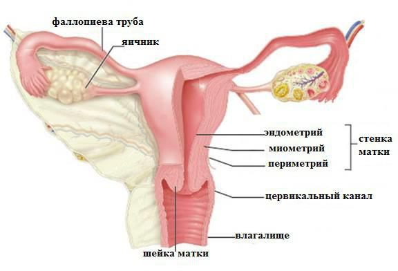
Bypassing the submucous base, the wall of the uterus after the myometrium passes into the endometrium - the mucous membrane. This is the inner layer, reaching a thickness of 3 mm. It has a longitudinal fold in the anterior and posterior region of the cervical canal, from which small palm-shaped branches depart at an acute angle to the right and left. The rest of the endometrium is smooth. The presence of folds protects the uterine cavity from the penetration of the vaginal contents, which are unfavorable for the internal organ. The uterine endometrium is prismatic, uterine tubular glands with vitreous mucus are located on its surface. The alkaline reaction they give preserves the viability of the sperm. During the period of ovulation, secretion increases and substances enter the cervical canal.
Uterine ligaments: anatomy, purpose
In the normal state of the female body, the uterus, ovaries and other adjacent organs are supported by the ligamentous apparatus, which form smooth muscle structures. The functioning of the internal reproductive organs largely depends on the condition of the muscles and fascia of the pelvic floor. The ligamentous apparatus consists of suspending, fixing and supporting. The set of performed properties of each of them ensures the normal physiological position of the uterus among other organs and the necessary mobility.
The composition of the ligamentous apparatus of internal reproductive organsApparatus | Functions Performed | Ligaments forming the apparatus |
Suspensory | Connects the uterus to the walls of the pelvis | Paired wide uterine |
Supporting ligaments of the ovary |
Own ligaments of the ovary |
Round ligaments of the uterus |
Latching | It fixes the position of the organ, stretches during pregnancy, providing the necessary mobility | The main ligament of the uterus |
Cystic uterine ligaments |
Sacro-uterine ligaments |
Supporting | Forms a pelvic floor, which is a support for the internal organs of the genitourinary system | Muscles and fascia of the perineum (outer, middle, inner layer) |
The anatomy of the uterus and appendages, as well as other organs of the female reproductive system, consists of developed muscle tissue and fascia, which play a significant role in the normal functioning of the entire reproductive system.
Suspension device characteristic
The paired ligaments of the uterus make up the hanging apparatus, thanks to which it “attaches” at a certain distance to the walls of the small pelvis. The wide uterine ligament is a fold of the peritoneum of the transverse type. It covers the body of the uterus and the fallopian tubes on both sides. For the latter, the ligament structure is an integral part of the serous cover and mesentery. At the side walls of the pelvis, it passes into the parietal peritoneum. The supporting ligament departs from each ovary, has a wide form. It is characterized by durability. Inside it passes the uterine artery.
Own ligaments of each of the ovaries originate from the fallopian fundus on the back side below the branch of the fallopian tubes and reach the ovaries. Inside them, the uterine arteries and veins pass, so the structures are quite dense and strong.
One of the longest hanging elements is the round ligament of the uterus. Its anatomy is as follows: the ligament has the appearance of a cord extending up to 12 cm. It originates in one of the corners of the uterus and passes under the front sheet of the broad ligament to the internal opening of the groin. After that, the ligaments branch into numerous structures in the fiber of the pubis and labia majora, forming a spindle. It is thanks to the round ligaments of the uterus that it has a physiological inclination anteriorly.
The structure and location of the fixative ligaments
The anatomy of the uterus was supposed to suggest its natural purpose - the bearing and birth of offspring. This process is inevitably accompanied by an active contraction, growth and movement of the genital organ. In this connection, it is necessary not only to fix the correct position of the uterus in the abdominal cavity, but also to provide it with the necessary mobility. Just for such purposes, fixing structures arose.
The main ligament of the uterus consists of plexuses of smooth muscle fibers and connective tissue radially spaced to each other. The plexus surrounds the cervix in the area of the internal pharynx. The ligament gradually passes into the pelvic fascia, thereby fixing the organ to the position of the pelvic floor. Bubble-uterine and pubic ligamentous structures begin at the bottom of the front of the uterus and attach to the bladder and pubis, respectively.
The sacro-uterine ligament is formed due to fibrous fibers and smooth muscles. It moves away from the back of the neck, envelops the rectum on the sides and connects to the pelvic fascia on the sacrum. In a standing position, they have a vertical direction and support the cervix.
Supporting apparatus: muscles and fascia
Uterine anatomy implies the concept of a “pelvic floor”. This is a combination of muscles and fascia of the perineum, which make up it and perform a function supporting a woman's internal genital organs . The pelvic floor consists of the outer, middle and inner layer. The composition and characteristics of the elements included in each of them are given in the table:
Female uterus anatomy - pelvic floor structureLayer | Muscle | Characteristic |
Outer | Sciatic-cavernous | Steam room, ranging from sciatic tubercles to the clitoris |
Bulbous spongy | Steam room, wraps around the entrance to the vagina, thereby allowing it to contract |
Outdoor | It compresses the anus with a “ring”, surrounds the entire lower part of the rectum |
Surface transverse | Weakly developed paired muscle. It comes from the sciatic tubercle from the inner surface and attaches to the perineal tendon, connecting with the same muscle going from the back |
Medium (urogenital diaphragm) | m. sphincter urethrae externum | Compresses the urethra |
Deep cross | The steam room is located between the symphysis, pubic and ischium. |
Internal (pelvic diaphragm) | Pubic-coccygeal | Paired branches m. levator ani, which raises the anus. Well developed. |
Iliac-coccygeal |
Sciatic-coccygeal |
The normal anatomy of the uterus and appendages is provided precisely due to the pelvic floor, which is the main support of the internal organs of the genitourinary system. The correct arrangement of organs is the key to their healthy functioning. Damage and significant weakening of the muscles of the pelvic floor threatens with prolapse and even prolapse of organs.
The structure of the ovaries and appendages
The anatomy of the uterus and ovaries is reproductive organs, interconnected by means of the fallopian tubes. The ovaries are the sex glands located on both sides of the uterus. Inside them, during the menstrual cycle, the eggs mature, which then enter the uterine cavity through the fallopian tubes.
The ovaries are fixed with a suspensory ligament and mesentery. Unlike the uterus, they are not covered by the peritoneum. The structure of the ovary is based on the brain and cortex. In the latter are mature follicles. On the inside, a granular layer adjoins the wall, in which the egg lies. It is surrounded by a radiant crown and a transparent zone.
During ovulation, the follicle approaches the outer layer of the ovary and bursts. So the egg is released and enters the uterine cavity with the help of the fallopian tube. A bursting follicle replaces the corpus luteum, which in the absence of pregnancy gradually disappears. If fertilization occurs, the corpus luteum continues to exist for the entire period to perform intracretory functions.
The surface of the ovaries is covered with a white membrane formed by connective tissue. Each ovary is surrounded by appendages having a convoluted shape and consisting of longitudinal and transverse tributaries. They are considered vestigial entities.
The fallopian tubes
A paired organ through which an egg from the abdominal cavity enters the uterus. The fallopian tubes look like oval ducts and extend in the upper part of the wide ligament of the uterus. Their length can be up to 13 centimeters, and a diameter of 3 mm. The egg is transported using the uterine and abdominal openings, the name of which corresponds to the cavities into which it exits.
Fallopian tubes consist of:
- uterine part - located in the thickness of the uterus;
- isthmus - the narrowest part with thick walls;
- ampoules;
- funnels - through their lumen the egg enters the fallopian tube;
- fringe - they direct the egg to the funnel.
Inside the tube there is a mucous membrane with ciliary epithelium and longitudinal folds, the number of which grows as you approach the abdominal opening. On the outside, the fallopian tubes are covered with a serous membrane.
The structure of the circulatory system
Blood supply to the genital organ occurs due to the uterine artery, which is a branch of the internal iliac artery. The anatomy of the uterus and fallopian tubes involves the outflow of blood from two sides, so the artery has two branches. Each of them is located along a wide ligament, then being divided into smaller vessels going to the front and back surfaces of the organ. Near the uterine fundus, the vessel branches again to provide blood flow to the fallopian tubes and ovaries.
The uterine veins form from the venous plexus, where venous blood flows. From here veins begin, which then flow into the internal iliac, ovarian veins and the plexus of the rectum. Venous outflow after the uterine and ovarian veins passes to the iliac and inferior vena cava.
The outflow of lymph from the internal genital organs
The lymph nodes to which lymph is directed from the body and cervix are the iliac, sacral and inguinal. They are located at the site of passage of the iliac arteries and on the front of the sacrum along the circular ligament. Lymphatic vessels located at the bottom of the uterus reach the lymph nodes of the lower back and inguinal region. The general plexus of the lymphatic vessels from the internal genital organs and rectum is located in the Douglas space.
Innervation of a woman’s uterus and other reproductive organs
The internal genital organs are innervated by the sympathetic and parasympathetic autonomic nervous system. Nerves to the uterus are usually sympathetic. On their way, spinal fibers and structures of the sacral nerve plexus join. The contractions of the uterine body are regulated by the nerves of the superior hypogastric plexus. The uterus itself is innervated by the branches of the uterine-vaginal plexus. The cervix usually receives impulses from the parasympathetic nerves. The ovaries, fallopian tubes and appendages are innervated from both the utero-vaginal and ovarian plexuses of the nerves.
Functional changes during the monthly cycle
The wall of the uterus is susceptible to changes both during pregnancy and throughout the menstrual cycle. The sexual cycle in the female body is characterized by a set of ongoing processes in the ovaries and uterine mucosa under the influence of hormones. It is divided into 3 stages: menstrual, postmenstrual and premenstrual.
Desquamation (menstrual phase) occurs if fertilization has not occurred during ovulation. The uterus, a structure whose anatomy consists of several layers, begins to reject the mucous membrane. A dead egg comes out with it.
After rejection of the functional layer, the uterus is covered only with a thin basal mucosa. Postmenstrual recovery begins. In the ovary, the corpus luteum is again produced and the period of active secretory activity of the ovaries begins. The mucous membrane thickens again, the uterus prepares to accept a fertilized egg.
The cycle continues uninterruptedly until fertilization occurs. When an embryo is implanted in the uterine cavity, pregnancy begins. With each week, it increases in size, reaching 20 or more centimeters in length. The birth process is accompanied by active contractions of the uterus, which contributes to inhibition of the fetus from the cavity and the return of its size to the antenatal.
The uterus, ovaries, fallopian tubes and appendages together form a complex system of female reproductive organs. Thanks to the pelvic floor and mesentery, the organs are firmly fixed in the abdominal cavity and are protected from excessive displacement and prolapse. A large uterine artery provides the bloodstream, and several nerve bundles innervate the organ.