As you know, for the normal functioning of the central nervous system, in particular the brain, the level of oxygen and the amount of glucose are extremely important. These substances are delivered to the nerve tissue along with blood. And the transport system in this case is the arteries of the brain. Today, many people are interested in more information about the blood supply to the brain. What vessels carry blood to the central nervous system? How is blood outflow carried out? What are the symptoms of impaired blood flow? What diagnostic measures are the most effective? What is the difference between CT and MRI of the brain? How to eliminate circulatory problems and is it possible to do it yourself? The answers to these questions will be interesting.
general information
For the normal functioning of the human brain, a sufficient amount of resources is needed. In particular, the central nervous system is extremely sensitive to oxygen and sugar levels in the blood. About 15% of all circulating blood passes through the vessels of the brain. On average, total brain blood flow is 50 ml of blood for every 100 g of brain tissue per minute.
There are four main cerebral arteries that fully satisfy the needs of this organ: two vertebrates and two internal carotid ones. Of course, it is worth considering the anatomical features of the body. What areas of blood supply to the brain exist? What happens when blood flow is disturbed?
Internal carotid arteries
These vessels are branches of the carotid artery (common). As you know, the common carotid arteries (right and left) are located in the lateral parts of the neck. If you put your fingers on the skin, then through the tissue you can easily feel the characteristic pulsation of the vascular walls. At about the level of the larynx, the common carotid artery branches into the external and internal. The inner penetrates through the hole in the skull, supplies blood to the tissues of the brain and eyeballs. The external carotid artery is responsible for the blood supply to the skin of the head and neck.
Vertebral Arteries
When examining the arteries of the brain, one cannot but mention the vertebral arteries. They branch off from the subclavian arteries, after which they pass through the holes of the transverse processes of the cervical vertebrae, and then penetrate the cranial cavity through the occipital foramen. It is worth noting that after entering the cranial cavity, the vessels connect to each other, forming a very specific arterial circle.
The connecting arteries of the willis circle are a kind of “security system”. If the blood flow in one of the vessels is disturbed, then due to the presence of the arterial circle, the load is redirected to other, healthy arteries. This helps to maintain blood circulation in the brain at the right level, even if one of the vessels is out of order.
Cerebral arteries
Cerebral arteries branch from the internal carotid artery. The anterior and middle vessels provide nutrition to the deep cerebral regions, as well as the surfaces of the cerebral hemispheres (internal and external). There are also posterior vertebral arteries, which are formed by branches from the vertebral arteries. These vessels carry blood to the cerebellum and brain stem. The large cerebral arteries diverge, forming a mass of small vessels that sink into the nerve tissues, providing them with food. According to statistics, cerebral hemorrhages in most cases are associated with a violation of the integrity of the above vessels.
What is the blood-brain barrier?
In modern medical practice, a term such as the blood-brain barrier is often used. This is a kind of system of transport and filtration of substances, which prevents the ingress of certain compounds from capillaries directly into nerve tissues. For example, substances such as salt, iodine, and antibiotics do not normally penetrate brain tissue. That is why, during the treatment of infections of the brain, antibacterial agents are injected directly into the cerebrospinal fluid - this way the antibiotic can penetrate the brain tissue.
On the other hand, alcohol, chloroform, morphine, and some other substances easily penetrate the blood-brain barrier, which explains their intense and almost instantaneous effect on brain tissue.
Carotid pool: features of anatomy
This term refers to the complex of the main carotid arteries, which originate in the chest cavity (including branches from the aorta). The carotid pool provides blood to most of the brain, skin and other structures of the head, as well as the visual organs. Violation of the functioning of the structures of this pool is dangerous not only for the nervous system, but also for the whole organism. Atherosclerosis most often causes blood circulation problems. This ailment is associated with the formation of a kind of plaque on the inner walls of the vessels. Against the background of atherosclerosis, the lumen of the vessel narrows, the pressure in it rises. The development of the disease is associated with a number of dangerous consequences, including embolism, ischemia and thrombosis. These pathologies in the absence of timely treatment can result in the death of the patient.
Vertebro-basilar system
In modern medical practice, a term such as the vertebro-basilar system, or the Zakharchenko circle, is often used. This is a complex of vertebral vessels. The structure also includes the basilar artery. Vertebral vessels, as already mentioned, originate in the chest cavity, and then pass through the channels of the cervical vertebrae and reach the cranial cavity. The basilar artery is an unpaired vessel that is formed by connecting the vertebral arteries. This part of the bloodstream provides nutrition to the posterior parts of the brain, including the cerebellum, the medulla oblongata, and part of the spinal cord.
Lesions of the above-described vessels (starting from mechanical injuries and ending with atherosclerosis) often end up with thrombosis. Violation of the blood supply to those brain structures that form this organ can lead to the appearance of various neurological symptoms and stroke.
Veins and blood outflow
Many people are interested in how the arteries and veins of the brain work. We have already examined the ways in which blood flows to the brain. As for the outflow system, it is carried out through the veins. The superior and inferior superficial veins collect blood from the subcortical layer of white matter and the cortical part of the cerebral hemispheres. Through the cerebral veins, blood is collected from the cerebral ventricles, internal capsule, and subcortical nuclei. All of the above vessels subsequently flow into the venous sinuses of the dura mater. From the sinuses, blood flows through the vertebral and jugular veins. With the external vessels, the sinuses communicate through the diploic and emissary veins. By the way, these vessels have some features. For example, veins that collect blood from brain structures are devoid of valves. A large number of vascular anastomoses are also observed.
Blood flow in the structures of the spinal cord
The spinal cord receives blood from the anterior, two posterior and radicular-spinal arteries. The back spinal vessels give rise to the vertebral (spinal) artery - they are directed along the dorsal surface of the spinal cord. The anterior spinal artery is also a branch of the vertebral vessels - it lies on the anterior spinal surface.
The above vessels only feed the first two or three cervical segments. Blood circulation of the rest of the spinal cord is due to the work of the radicular-spinal arteries. In turn, these vessels, which go down and go along the entire spine, receive blood due to communication with the ascending cervical, intercostal and lumbar arteries. It is also worth saying that the spinal cord has a highly developed system of veins. Small vessels take blood directly from the tissues of the spinal cord, and then flow into the main venous channels that run along the entire spine. From above, they connect to the veins of the base of the skull.
Cerebrovascular accident
Considering the arteries of the brain, one can not but mention the pathologies that are associated with circulatory disorders. As already mentioned, the human brain is extremely sensitive to oxygen and blood sugar, so a deficiency of these two components negatively affects the functioning of the whole organism. Prolonged hypoxia (oxygen starvation) leads to the death of neurons. The result of a sharp decrease in glucose levels is loss of consciousness, coma, and sometimes death.
That is why the circulatory system of the brain is equipped with a kind of protective mechanisms. For example, the venous system is rich in anastomoses. If the outflow of blood through one vessel is impaired, then it moves differently. The same applies to the Willis circle: if the current along one artery is disturbed, its functions are taken over by other vessels. It is proved that even if the two components of the arterial circle do not work, the brain still gets enough oxygen and nutrients.
But even such a well-coordinated mechanism sometimes fails. Pathologies of cerebral vessels are dangerous, therefore it is important to diagnose them in time. Frequent headaches, periodic dizziness, chronic fatigue - these are the first symptoms of cerebrovascular accident. If untreated, the disease can progress. In such cases, chronic cerebrovascular accident, dyscirculatory encephalopathy develops. Over time, this ailment does not disappear - the situation only worsens. Lack of oxygen and nutrients leads to slow death of neurons.
This, of course, affects the work of the whole organism. Many patients complain not only of migraines and fatigue, but also of tinnitus, periodically occurring pain in the eyes (for no apparent reason). The occurrence of mental disorders and memory impairment. Sometimes nausea, the appearance of tingling on the skin, numbness of the limbs. If we talk about acute violation of cerebral circulation, then, as a rule, it ends with a stroke. This condition rarely develops - the heartbeat becomes more frequent, consciousness gets confused. There are problems with coordination, there are problems with speech, divergent strabismus, paresis and paralysis (usually unilateral) develop.
As for the reasons, in most cases, blood flow disorders are associated with atherosclerosis or chronic hypertension. Risk factors include diseases of the spine, in particular osteochondrosis. Deformation of the intervertebral discs often leads to displacement and compression of the vertebral artery, which feeds the brain. If you notice any of the above symptoms, consult a doctor immediately. If we are talking about acute circulatory failure, the patient needs immediate medical attention. Even a few minutes of delay can harm the brain and lead to a host of complications.
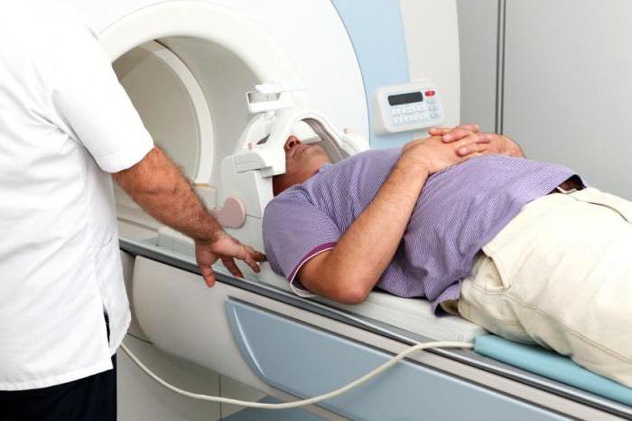
CT and MRI of the brain
The price in Moscow (as in any other city) for such procedures is quite high. Therefore, many people are interested in additional information about such diagnostic measures. These procedures are considered the most informative. So what is the difference between CT and MRI of the brain? In fact, the goal of such procedures is one and the same - scanning the human body with the further construction of the body image "in section".
Nevertheless, the operation scheme of the devices themselves is different. The basis of the work of ART equipment is a feature of the behavior of a hydrogen atom in a powerful magnetic field. But with computed tomography, special detectors receive information about tissues and organs, which capture radio emission that has passed through the human body through x-ray tubes. Both devices transmit all the data to a computer that analyzes the information, forming pictures.
How much does a brain MRI? Prices in Moscow vary depending on the policy of the selected clinic. The study of cerebral vessels will cost about 3,500-4,000 rubles. The cost of CT is slightly lower - from 2500 rubles.
By the way, these are not the only diagnostic measures that help diagnose certain blood flow disorders. For example, angiography of the arteries of the brain provides a lot of useful information. The procedure is carried out by introducing into the vessels a special contrast agent, the movement of which is then tracked using x-ray equipment.
What medications are prescribed to improve blood circulation in the brain? Drugs and the right diet
Unfortunately, many people are faced with a problem such as impaired blood flow in the vessels of the brain. What to do in such cases? What medications are prescribed to improve blood circulation in the brain? The drugs, of course, are selected by the attending physician, and experimenting with such agents on their own is not recommended.
As a rule, medications that prevent platelet aggregation and blood coagulation are included in the treatment regimen. Vasodilator drugs have a positive effect on the state of nerve tissues. Nootropic drugs also help improve blood circulation and, accordingly, trophic tissue. If there are indications, the doctor may prescribe the use of psychostimulants.
People at risk are advised to reconsider their lifestyle and, above all, nutrition. Experts advise to include in the menu vegetable oils (linseed, pumpkin, olive), fish, seafood, berries (cranberries, lingonberries), nuts, sunflower and flax seeds, dark chocolate. It is proved that regular use of tea has a positive effect on the circulatory system.
It is important to avoid physical inactivity. Feasible and regular physical activity enhances blood flow to tissues, including nerve. Positive effect on the circulatory system is the sauna and bath (in the absence of contraindications). Of course, in the presence of any disorders and disturbing symptoms, you should consult a doctor and undergo a medical examination.