In medical practice, there are a large number of analyzes, laboratory tests and various methods for determining the condition of internal organs and our body in general. One such research method is kidney urography.
What is urography?
To begin with, the word "urography" itself consists of two concepts that answer the question posed. The first word is “ur”, “urine” is urine, the second is “grafia” - to depict, write. This is such an x-ray method for examining the urinary system, in which its condition is evaluated. There are many diseases of the excretory system , therefore, excretory urography of the kidneys shows what abnormalities are present in each specific case.
Indications for the study
In order to verify the damage to the kidneys or urinary tract, doctors prescribe an examination. Most often, it is done with suspicion of the following diseases:
- abnormalities in the development of the ureters and kidneys;
- hematuria of unknown etiology;
- stones in the ureters and kidneys;
- private kidney diseases such as pyelonephritis, hydronephrosis, tuberculosis, glomerulonephritis, nephrogenic hypertension and others;
- kidney tumors ;
- kidney injury.
Also, with the help of a study such as urography of the kidneys, you can see the condition of the abdominal cavity to examine the state of the digestive system.
Contraindications to the procedure
As in other cases, this examination has a negative effect on some people. Excretory urography of the kidneys is not recommended for patients with pathologies such as renal and liver failure. A study is contraindicated for patients who have had a stroke and myocardial infarction. A separate group of non-admission to the procedure are pregnant women. Kidney urography during pregnancy is contraindicated for women, since transmitted rays can cause the development of anomalies in the child.
Very rarely there are complications after the study, such as urticaria, anaphylactic shock and laryngospasm, this should be taken into account when prescribing urography for allergy sufferers.
How is the study?
When conducting this study, a
radiopaque substance is administered intravenously to a person
. In this case, the patient should be in a horizontal position. For urography, a special substance is used - sergazine, in a dosage of 40 ml per 40% solution, cardiotrust is also often used (in 35% solution 20 ml) or some other contrast agent. The drug is slowly injected into a vein, then 3-5 pictures are taken in the urinary tract and kidneys. To do this, the
x-ray tube should be located exactly in the middle between the navel and the pubis. You can start taking the first picture as early as 10 minutes after an intravenous injection. Then the specialist looks at the dynamics of the image on the screen. If the picture is good, pictures can be taken after 20 minutes, 40, 60, and also after 2 hours.
What does overview urography of the kidneys show?
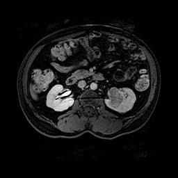
The study is very informative for the doctor. When conducting such studies, x-rays pass through the structural elements of the body. Using electromagnetic radiation, a negative image appears on a special plate. Here, the specialist looks at the density of the tissue or organ - the lighter the picture on the film, the higher it is. Kidney urography allows you to study their excretory ability. In addition, the sizes of morphological elements are clearly visible here: calyx, pelvis. The film shows whether there are stones where they are located, whether they managed to cause changes in the ureters and kidneys. Also, with the help of this study, hydronephrosis, various kinds of tumors, and tuberculosis of the kidneys can be determined. Urography informatively displays the processes and functions of a healthy kidney, if the second is damaged. After the study, the specialist compares the results of urography and other clinical data. If the kidneys function normally, there is good urodynamics, then the study may end in 30 minutes. However, there are cases when it is delayed up to 24 hours after the introduction of a special substance.
For additional study of the reserve capabilities of the kidneys on the basis of the introduced radiopaque substance, furosemide (40 mg) is administered after 20 minutes. This special technique, called pharmacouurography, allows you to determine the dilatation of the upper urinary tract, that is, latent disorders provoked by complex diseases. For this, furosemide must be dissolved in an isotonic sterile solution of sodium chloride.
Kidney urography: preparing for it
Before the study, it is important to carefully consider the doctor's recommendations. Most often, the preparation procedure is the same for everyone, of course, there are exceptions. Early in the morning, before the event, the patient eats fried white bread, as well as meat. After this, a person is given a cleansing enema. No other special preparatory actions are required.
Before the procedure, you must discuss with the doctor all the exciting issues. For example, how the rays affect the human body, or how many x-ray studies in a certain period of time are safe for the body. Some even keep records of such studies in order to provide the specialist with accurate information about themselves. Be sure to inform about the presence of pregnancy in you or suspicion that it may be.
Kidney urography in children is carried out without any special differences from the usual adult study. But there are some nuances. It is very important to correctly determine the dose of contrast medium. It depends on the age of the child, his body weight, the functional state of the liver and kidneys. There are distinctions by age for children: from several months to one year, 3 or 4 ml of a substance is prescribed, and from a year to three years - 2-3 ml. Since small children cannot withstand the study for a long time, for them, urography is carried out with minimal time ranges: from 1 minute to 40 - this is for children up to three years. For older, this range stretches to 60 minutes.