The cardiovascular system, by providing a constant flow of blood every second, supplies oxygen and nutrients to all the internal organs of a person, and therefore its value is undeniably high. And that is why, when the slightest disturbances occur in it, cascading failure reactions are caused in all other systems, and therefore symptoms always appear. But how is the examination of the heart and blood vessels performed? There are many methods for this.
Inspection
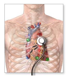
When the patient first visits the therapist for either a prophylactic purpose (physical examination) or specific complaints, the specialist must necessarily examine the area of the heart and conduct simple studies of this organ and its branches. So, first of all, the doctor conducts a general examination of the patient, paying attention to his skin (with diseases of this system, pallor and even cyanosis, dense cold edema, minor hemorrhages are possible), the condition of visible mucous membranes (sclera injection, white plaque at the root of the tongue) the development of the musculoskeletal system (hypotension, weakness, dystrophicity, or, conversely, obesity), the nature of the pulse (its presence and synchronism on both hands, holding the pulse in the cervical veins). Further, the doctor must conduct such a heart examination as percussion of its borders, which can reveal hypertrophy of individual chambers. Be sure to carry out his auscultation with the calculation of the number of heart contractions, detailing its tones, rhythm, possible pathological noises.
Anamnesis
Finally, blood pressure is measured because it is an important indicator of the state of the cardiovascular system. Further, the doctor will necessarily detail the complaints, because a full examination of the heart includes a detailed history. So, diseases of the cardiovascular system are characterized by chest pains (often of a pressing, constricting nature) or, more precisely, behind the sternum, shortness of breath (appears with increased physical exertion, normal, and with pathology with a slight exertion, or even at rest),
heart palpitations and a feeling of any “interruptions” in the work of the heart, manifestations of high blood pressure (headaches, dizziness, heaviness in the body). Be sure to find out the time of their appearance, the factors that provoke and eliminate them, the intensity.
Other important aspects
Also, a heart examination includes the fact that the patient is asked what he associates with the development of his disease, thereby identifying risk factors. So, it can be a strong emotional shock the day before (death of a loved one, stress at work), weight lifting or the performance of intolerable physical work. Symptoms also appear with changing weather conditions. An important criterion is heredity, because most diseases (diabetes mellitus, arterial hypertension,
coronary heart disease) are prone to transmission to the next generation. As a rule, a correctly collected history gives 50% of the patient's clinical diagnosis. After talking with the patient and conducting his examination, the doctor must send his ward to a heart examination. We should recall the anatomy and physiology of this organ.
A little bit about a normal heart
So, it is, roughly speaking, a pump, consisting mainly of muscles and a complex system of blood vessels. Inside it there are four chambers that communicate with each other in a strictly defined way and ensure the constant movement of blood. And in order for the heart itself to continuously contract and relax, in its tissues there are conductor structures along which a nerve impulse passes, thereby causing alternating muscle tension in each chamber and opening-closing valves between them. Therefore, all methods of examining the heart can be aimed either at visualizing the anatomy of this organ (ultrasound, Doppler mapping, computed tomography, chest x-ray, radioisotope methods) and directly arteries and veins (sounding of the great vessels, angiography, coronarography), or at the study of the condition its conductive system (electrocardiography, bicycle ergometry), or the audio of its tones and noises (phonocardiography).
Echocardiography
As you can see, the examination of the heart must be certainly detailed, detailed, not losing sight of anything. Because the defeat of the cardiovascular system can be both a manifestation of an independent disease, and a consequence of the pathology of another system. If we talk about visual
diagnostic methods, then the first to come to mind is Echo-KG or, as it is also called,
ultrasound of the heart. What the device shows during this important study is logical to guess. By ultrasound penetrating deep into the tissues and returning them back to the screen, an image appears that allows you to evaluate the structure of the heart, the size of its cavities, the condition of the valves and main vessels. In addition, this method is non-invasive and takes place without radiation, and therefore it can be used even by pregnant, lactating and children. Although more effective
computed tomography still cannot displace ultrasound from diagnosis.
Ultrasound Benefits
At different gestational periods, a woman periodically undergoes an ultrasound scan of the heart for the fetus, which shows an unclosed arterial duct, stenosis of the mouth of the vessels, prolapse or insufficiency of valves, the state of the interventricular and atrial septum, and other congenital malformations. Another important advantage of this method for the patient himself and the medical institution is its relative cheapness, the possibility of outpatient testing, the short duration of the study, as well as the instant receipt of a snapshot and interpretation of all data. And therefore it is so popular in the use for the diagnosis of ultrasound of the heart.
What does the study of blood vessels
In obese people, as well as patients with diabetes mellitus, the most common lesions of the cardiovascular system are atherosclerotic lesions of blood vessels, as well as hyalinosis of their walls. Therefore, it is so necessary to examine the blood vessels of the heart, because only they nourish this important organ, and its work requires a tremendous amount of energy and nutrient substrates. So, first a catheter is inserted into the femoral or subclavian artery , through which the contrast medium, clearly visible on the X-ray screen, fills the vessels. The most important method for atherosclerosis, coronary heart disease, myocardial infarction is a coronary examination of the heart vessels. It reveals their patency, the correctness of their course. Also under his control, many operations are carried out on this important organ.
Summary
Thus, at present there are many methods for studying cardiac and vascular pathology, however, each of them has strict indications and contraindications, and therefore it is economically unrealistic and diagnostic sense to carry them out to everyone and everyone. Therefore, the key link is precisely a competent doctor who carefully procures the patient and prescribes the necessary treatment for him or sends him to a more competent institution.