The human body is pierced from head to foot with blood vessels. They allow the body to function normally and carry nutrients and oxygen throughout the body. Among them there are also vessels that play a vital role for humans.
Carotid artery
Each of us at least once in our life damaged some part of the body, for example, when a finger was cut, blood began to flow out of it. It is not difficult to stop such bleeding, since the diameter of the vessel is quite small and the pressure in it is small. In addition, there are platelets in human blood that clog the cut, and after a couple of minutes the blood itself stops flowing.
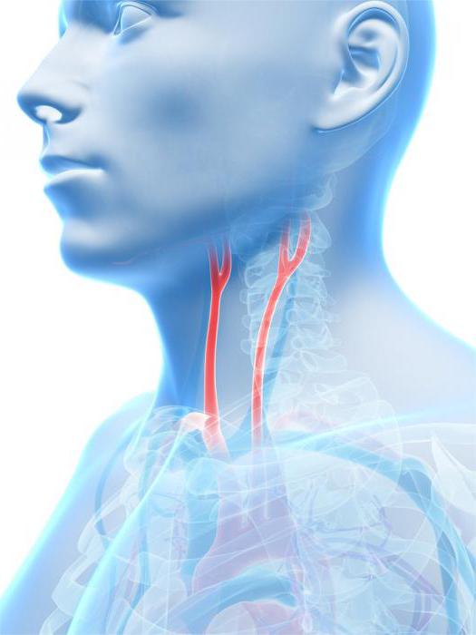
But this does not always happen: in the human body there are vessels that differ in large diameter and blood pressure, which moves through them. Usually they are the most important in the human body, and their damage and lack of medical care can lead to serious blood loss. One of these is the carotid artery.
This blood vessel is a paired artery that begins in the chest and branches out, heading toward the head. Because of this, its main functions can be considered the blood supply to the brain, eyes and other parts of the human head.
More about the structure of the carotid artery and its functions
The carotid artery has two branches: the right and left. The first originates in the area of the brachial trunk. The left artery, in turn, begins in the area of the aortic arch. Due to such anatomical features, the left artery is a couple of centimeters longer than the right. Then it vertically moves upward, located in the neck, then branches and is located in different parts of the head.
The main function of this artery is the blood supply to the brain. This can happen only when this vessel does not have pathologies and various diseases that interfere with normal blood circulation. When a blockage in the arteries occurs, a person will most likely need surgery.
External carotid artery
This kind of artery is considered one of the main components of one common trunk of the carotid artery. It starts from a single artery, located at the level of the carotid triangle, one of its parts. First, it passes closer to the middle of the artery located inside, and then much lateral relative to it.
Initially, this artery is covered with muscle, and if we consider its location in the region of the carotid triangle, then you can observe it under the subcutaneous muscle located in the neck. The artery does not end there, its division occurs. In the region of the lower jaw, approximately at the level of the neck, the first branches of the external carotid artery appear. They are represented by the maxillary and superficial temporal artery. Then other branches of the external carotid artery appear, they diverge in different directions in the corresponding directions. Therefore, the anterior, middle, and posterior branches of the external carotid artery are determined here. Each of them is responsible for the normal functioning of certain parts of the human body, supplying them with nutrients and oxygen.
Front group
It is these areas related to the external branch of the trunk of the carotid artery that include quite impressive vessels. The peculiarity of this group is that it allows blood to flow to organs located in the face and throat. Therefore, the functioning of the larynx, face, tongue, thyroid gland depends on their normal operation. From the common vessel, which is a branch of the external carotid artery, three main vessels depart, rather large in size. Then there is another division into smaller vessels, this differentiation allows you to apply blood to all necessary parts of the body.
The front group of branches of the external carotid artery includes three main vessels, each of them has a specific function and location.
Superior thyroid artery
Its branch occurs at the level of the horns at the very beginning of the hyoid bone. This arrangement allows this particular artery to supply blood to the thyroid gland and, of course, to the parathyroid. Also, thanks to this artery, blood enters the larynx, passing through the superior artery in the area of the mastoid muscle.
After that, it, like most vessels in the human body, is again divided. And in the upper thyroid artery, the hyoid and cricoid branches appear. One of them, namely the hyoid, becomes the main vessel that nourishes the nearest muscles, and the hyoid bone.
As for the cricoid branch, it allows blood to flow to the corresponding muscle. After this, it connects to a vessel similar to it on the other hand.
The superior laryngeal artery allows blood to be delivered to the epiglottis and larynx. With its help, it seems possible to enrich the shells of these organs with oxygen, as well as the muscles located around.
Lingual artery
This vessel, like the previous ones, is a component of the branch of the external carotid artery, a branch occurs just above one of the vessels, in particular, the thyroid. This happens in the region of the hyoid bone, then it moves and gradually reaches the region of the Pirogov triangle. Then the lingual artery goes to the point from which it got its name, that is, to the language itself, it is located below. Although. Compared to other arteries, the lingual is considered not so big, it also has its own smaller vessels.
For example, the deep artery of the tongue looks like a large branch of the lingual artery. Its location is quite interesting: first it rises up and reaches the so-called base of the language. Then it continues to move along it and reaches the very tip. This vessel surrounds several muscles, in particular, the lingual and lower longitudinal.
In addition, there is a suprahyoid branch, its main function remains the blood supply to the hyoid bone. Accordingly, it is located on the upper edge of this bone. The hyoid artery is located in the region of the hyoid muscle, directly above it. Its functional features are in the blood supply to part of the oral cavity, thanks to it oxygen is supplied to all components of the human oral cavity. This includes the mucous membrane of the mouth, salivary glands, and even the gums. The dorsal branches have a peculiar arrangement, so they can be observed in the area of one of the muscles, in this case the hyoid.
Facial artery
This type of vessel branches in the region of the corner of the lower jaw, and then goes through the gland located near, that is, the submandibular. This vessel is not for nothing called the facial artery, since starting from the neck, it goes through the region of the lower jaw, gradually moving to the area of the face. Then it goes forward and moves to the top. The ends of the vessels end at the corners of the mouth, and another branch reaches the eyes. In addition, the artery itself includes additional vessels, respectively, other branches appear.
Despite the fact that there are mainly branches of the external carotid artery on the neck, the smaller arteries that make up the group are located in the face and part of the person’s mouth. The amygdala branch goes to the palatine tonsil, and from branching it goes through the sky. She also goes to the base of the tongue, reaching there along the wall of the human oral cavity.
As for the palatine artery, its location is directly from the very base of the facial artery, which is part of a group called the anterior branches of the external carotid artery. The ascending palatine artery ends in the area of the pharynx, in particular, its mucosa and, in addition, the tonsil. The last branches also reach the pipes responsible for normal hearing.
The chin artery goes through the hyoid muscle, to be more precise, through the outer surface of this muscle. The ends of the vessel move to the area of the chin and certain cervical muscles.
Back group
The posterior branch of the external carotid artery, like the previous ones, has its own branches of blood vessels. The auricle departs from it, and in this place the occipital artery originates. With their help, blood supply to the visible inner part of the ear occurs. In addition, thanks to these arteries, blood flows to the muscles of the neck located behind, in the back of the head, as well as the channel of the facial nerve. A distinctive feature of this branch is that it has the ability to penetrate the membrane of the brain.
Occipital artery
Departs separately, is almost as high as the front. Its location - in the area of the biceps muscle, is located under it, after which there is a movement into the furrow near the temple. Further, its path passes under the skin, where it is located, the back of the head is involved, and the branching occurs in the epidermis of the occipital region.
After going all this way, they are connected with the same branches that go from the opposite side. Connection is also made with other branches, some vessels of the spinal column.
The occipital artery has a division into several smaller vessels, respectively, appear auricular, descending, mastoid branches. The first goes straight to the visible inner part of the human ear, and after passing it, it becomes one with the other branches of the posterior ear artery. The descending reaches the most hidden corners, as it goes to the area of the neck that is further than the rest. As for the mastoid, it lies in the shell of the human brain, in the corresponding channels available there.
Posterior ear artery
The branches of the external and internal carotid arteries play an important role in the human body, as well as their smallest branches. For example, this vessel is directed obliquely backward, it goes from the biceps muscle, then spreads in this way: it passes from the edge of the posterior abdomen. It is also divided into three smaller branches. One of these vessels will be the occipital branch.
Its location corresponds to the base of the
mastoid process, allows blood to enter the skin located in the occipital region. The auricular branch paved its way along the back in the ear region and allows blood to be supplied with blood to the visible parts of the inside of the human ear. The styloid artery plays an equally important role: the facial nerve depends to a large extent on its normal operation, because it is precisely to it that blood flows, the location partially corresponds to the temporal bone.
Middle group
The middle group of branches of the external carotid artery has fewer branches in comparison with the previous ones. In fact, this group includes one artery, which then branches into a number of smaller vessels, but its significance does not decrease from this.
The medial branches of the external carotid artery include the pharyngeal ascending artery and other vessels, which make it possible to supply nutrients, and most importantly, oxygen, to those muscles that are located on the face, that is, nourish the lips, cheeks, etc.
Ascending pharyngeal artery
After its branch, this artery takes a direction in the direction of the pharynx and passes along its wall. The branching of this vessel occurs so that the posterior meningeal artery goes towards the tympanic part and spreads further through the tympanic tubule, located in one of its cavities, in this case the lower one.
End branches
The terminal branches of the external carotid artery are a small number of blood vessels that are part of the carotid artery. This branch has two arteries, namely the maxillary and superficial temporal. They differ in size, and other vessels extending from them allow you to transfer blood to distant parts of the body.
Superficial temporal artery
This vessel is considered a continuation of the external carotid artery. Its passage corresponds to the visible surface of the inner part of the ear, namely its front wall, the artery is located under the skin. The movement goes up and goes towards the temple area. If you need to feel the pulsation, indicate the branches of the external carotid artery in this place. Here, determining the beating of the blood flow is quite simple.
Then another division occurs: the parietal, as well as the frontal artery appears. This happens at the level of the corner of the eye located near the temporal region. These arteries carry blood to the forehead, crown, and cranial muscle.
The final branches of the external carotid artery include a surface vessel, which is divided into five smaller ones. One of them is the transverse facial artery. This blood vessel is located in the area of the parotid gland, its duct. Then it moves towards the cheek and is located in the skin. Vessels spread in the infraorbital region and reach another type of muscle tissue - mimic.
The zygomaticoorbital allows blood to flow to some muscles of the eye, passing through the small zygomatic arch. The front auricles go to the ear, namely its visible surface of the inner part, there is also a middle temporal artery and branches located in the area of the gland located here.
The maxillary artery does not go with one trunk and is also divided into other vessels, in this case there are several sections, one of which is the jaw. It includes the smaller vessels departing from it, for example, it is a deep ear artery. There is also a fairly large artery called the lower alveolar. The most dense among the vessels of this group is the middle meningal, located in the direction of the lining of the brain.
Conclusion
The above information shows what the external carotid artery is. The topography of the branch divides it into 4 groups. All of them are important for a person, and a malfunction in one of them can affect not only problems in the area of a certain part of the body, but also the work of the whole organism. An important role is also played by small vessels, which extend from each branch, since they allow you to supply blood to the eyes, cheeks, chin, different parts of the head, pass both in the muscles and are located closer to the epithelium.