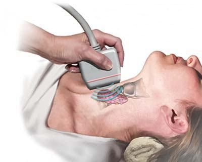
Today, ultrasound of the thyroid gland by many experts is considered the most informative method of studying the state of this organ. Moreover, ultrasound diagnostics is one of the most affordable today. Its undoubted advantage should be considered the ability to examine patients at any age.
What is research necessary for?
Ultrasound of the thyroid gland (the norm for healthy people will be given below) can be used for preventive examinations and medical examinations. Timely implementation of ultrasound often reveals organ defects, tumor changes, minimal foci of inflammation. However, at the same time, it is not possible to identify the very reason for the occurrence of violations using only this method. During the study, the specialist also studies the structure of paired formations - parathyroid glands. They are located in the left and right lobes of the organ. The lymph nodes located on the front of the neck of the person are also examined. As a matter of fact, ultrasound of the thyroid gland, the volume norm of which is different for women and men, is considered the first additional diagnostic method prescribed by the endocrinologist. In accordance with the data obtained, correction of the patient examination scheme can be carried out.
What is specifically studied during ultrasound?
When performing an ultrasound of the thyroid gland, the norm and deviations are estimated by several parameters. First of all, the structure of the organ is studied. In this case, the ability to reflect the sensor signal in the thyroid gland and salivary parotid gland is compared. The study allows you to study the echogenicity of the organ. This parameter indicates uniformity of tissue. As mentioned above, the parathyroid glands and lymph nodes are examined. In addition, the state of large vessels that are located near the organ is assessed. In particular, the jugular veins and the external carotid artery are examined . We study the volume of the organ, as well as the structure of the isthmus, uniting the lobes, the size of the thyroid gland. The norm of linear values depends on the age and gender of the patient. If necessary, an examination of other anatomical structures can be carried out: soft tissue of the neck, larynx and others.
Ultrasound of the thyroid gland. Decryption
The norm of volume in men is up to 25 ml, in women - up to 18. The description of the conclusion may look as follows: "The location of the organ is correct, the shape is normal, the contours are clear, even, there are no nodes, the echostructure is not changed, it is homogeneous. The lymph nodes of the subclavian, submandibular region are not enlarged." However, with certain pathologies, the size of the thyroid gland by ultrasound does not deviate from generally accepted parameters. Such diseases, in particular, include diffuse toxic goiter.
Pathologies that are detected by ultrasound
If you suspect which diseases, an ultrasound of the thyroid gland is prescribed? Dimensions, the norm of which is individual for each person, may indicate the presence or absence of thyroiditis. In the structure of the organ, seals, diffuse or local changes can be detected. In the latter case, small compacted nodes of various sizes are detected. In some cases, during a routine examination by an endocrinologist, they are not noticeable. In this regard, the doctor (to clarify the diagnosis) prescribes an ultrasound of the thyroid gland, the norm of the volume and linear parameters of which is indicated above.
Tumor Diagnosis
In most cases, during the study, a specialist can identify and distinguish between benign and malignant neoplasms in the thyroid gland. The latter are characterized by reduced echogenicity, the presence of calcium salts in the tissue, heterogeneity of the structure. The neoplasm can be of different sizes, including very small. After removal of the tumor, an ultrasound of the thyroid gland is re-prescribed. The norm will indicate the effectiveness of the measures taken. The study is recommended to be carried out regularly to exclude relapse.
When is an examination necessary?
Who is prescribed for thyroid ultrasound? How to prepare for the study? First of all, screening is necessary for people at risk. These include, in particular, people over forty years old, since it is at this age that the likelihood of neoplasms of a benign or malignant nature increases. The examination is necessary for patients working in hazardous industries, spending a lot of time at the computer, often in stressful conditions. Ultrasound is recommended for patients who have been prescribed hormonal drugs throughout their lives in connection with certain pathologies. Adverse heredity is also an indication for the purpose of the study. It is necessary to conduct a survey of pregnant women.
Ultrasound is recommended in this case, both at the planning stage, and throughout the term for any deviations.
Who else is prescribed an examination?
Diagnosis is recommended for people who have symptoms of thyroid pathologies. In particular, with unclear weight fluctuations, changes in the frequency of heart contraction, unexplained irritability or lethargy, which are not provoked by the use of medications or by violations of thermoregulation. In addition, ultrasound is recommended if thyroid hormone levels are reduced or increased, the norm for total thyroxin is 60.0-160.0 nmol / liter, and for T3 (free) 1.2-2.8 mIu / liter. If there are deviations, then an additional study will clarify the diagnosis. Before conducting an ultrasound examination, the patient does not need special training.
Additional features for ultrasound
When detecting autologous changes, a specialist can recommend an ultrasound scan with a CDK (digital Doppler mapping). This research method allows not only to study the features of the structure and structure of the organ, but also to evaluate the nature of interstitial blood flow. Based on all the data, a more accurate diagnosis is made. Especially often, CDC is used when tumor nodes are detected in the gland. When examining the features of blood flow, a specialist has the opportunity to understand the true causes of the development of pathology, the likelihood and direction of metastases against a malignant process. Under the control of ultrasound, a fine-needle biopsy of tissues from pathological lesions detected during a preliminary examination is performed.
Features of diffuse changes detected by ultrasound
These disorders, as a rule, provoke inflammatory processes in the thyroid gland. These include, in particular, chronic thyroiditis. During the examination, a decreased echogenicity of the organ, an increase in it in all directions, is noted. Typical features are diffuse tissue heterogeneity. With thyroiditis, several fuzzy delimited nodes can be detected. Their internal structure is similar to the structure of the surrounding tissue. When a large node is detected, the shape of the gland changes (it becomes diffuse-nodular).
Differential diagnosis
Ultrasound can distinguish multinodular goiter from chronic thyroiditis. This is especially important, since the choice of therapeutic measures will also depend on the diagnosis. So, autoimmune thyroiditis is treated conservatively, and multinodular goiter is treated surgically. Diffuse changes can accompany bazedovo disease (diffuse toxic goiter). They appear in the form of a uniform increase in the thyroid gland, in some cases by 2-3 times in comparison with the norm. However, in many cases, the severity of the pathology does not affect the size of the thyroid gland. The norm, as mentioned above, is individual for each patient. With pronounced manifestations of thyrotoxicosis in men, for example, a slight deviation from generally accepted parameters is noted. As a rule, the structure of the tissue is homogeneous, may be slightly dense, echogenicity is increased. In some cases, against the background of the considered changes, secondary nodules, accumulations of calcium salts and cysts can be detected.
Conclusion
Unfortunately, not everyone is able to notice metabolic disturbances. Most people realize that they are sick when they have serious problems with appetite or weight. The tendency to consume a large amount of sweet, frequent mood swings, hair loss - all these are signals about the presence of any disorders in the body. With such manifestations, you should consult a specialist. An endocrinologist will conduct an examination and prescribe the necessary tests and studies, including an ultrasound of the thyroid gland. Where to do an ultrasound scan? It is held in special rooms in which the appropriate equipment is installed. Diagnosis is carried out by a specialist - a uzist doctor. Today, ultrasound, as indicated above, is the most informative diagnostic method. This mainly explains its popularity. In addition, the study is available to a large mass of the population. The cost of the examination is much lower, and its information content is higher than that of thyroid radiography. Moreover, when conducting ultrasound, there is no radiation load on the patient's body. The undoubted advantage of the method is the possibility of its repeated use by patients at any age, including newborns, as well as pregnant women. Before conducting the study, the doctor must be informed of all the drugs taken by the patient, including vitamins. However, despite all the informativeness of the ultrasound examination, to make a diagnosis, taking into account only the results of ultrasound of the thyroid gland, will be incorrect. Of great importance is the whole clinical picture, information on the medical history. The results of other studies are also taken into account, including indicators of thyroid hormones (the norm for them is indicated above). Only on the basis of an assessment of all the data obtained in the complex during an ultrasound examination, the doctor can make a conclusion about the presence or absence of pathological changes and make an accurate diagnosis, according to which treatment will be prescribed.