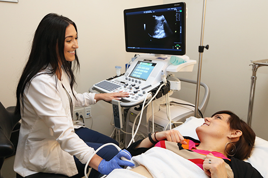
Ultrasound examination is currently considered one of the most informative and safe diagnostic methods. Ultrasound of the digestive tract allows you to evaluate the size and shape of the organs of the abdominal cavity and intestines, to analyze their structure and echogenicity, so that the doctor can identify abnormalities, make a diagnosis and prescribe adequate treatment.
The advantage of this method is its painlessness, accessibility, informational content and high speed of execution - from 20 minutes to half an hour.
Preparation for an ultrasound of the digestive tract: a study for adults
Most often, with the help of ultrasound, the state of such organs as the liver, kidneys, pancreas, gall bladder and spleen is assessed. Due to the accumulation of gases, the intestines are usually examined using other methods, but sometimes the doctor may prescribe an ultrasound.
Indications for it are complaints of poor health and pain in the abdomen, as well as various dyspeptic symptoms, including heaviness in the abdomen and increased gas production in people who do not use foods that cause flatulence.
Diet in preparation for ultrasound
It implies preparation for the study and diet before an ultrasound of the gastrointestinal tract. Three days before the diagnostic procedure, you need to make adjustments to your diet to eliminate bloating and flatulence. For this reason, you can not use:
- wheat and rye bread, any pastry;
- all kinds of cabbage;
- beans, peas and other legumes;
- sweet soda and regular mineral water;
- whole milk (especially if there is lactose intolerance);
- raw vegetables and fruits.
Featured Food
When preparing for an ultrasound of the digestive tract, it is recommended to eat lean meat and fish, best boiled or steamed, rice and oatmeal porridge in the water, baked apples, vegetable puree soups (without cabbage, peas and other prohibited vegetables).
Food should be fractional. In preparation for the study of ultrasound of the gastrointestinal tract per day, you need to drink 1.5 liters of water without gas, and you will have to refuse coffee and tea, as well as nicotine and chewing gum.
Other preparation features
For several hours on the eve of an ultrasound, you need to refrain from taking such drugs as aspirin, analgin and No-Shpa.
If the patient has difficulty emptying the bowel, preventive measures should be taken. 12 hours before the study, you need to drink a laxative or put a rectal suppository. But if this does not help, an enema is recommended. In any case, the intestines need to be empty.
In addition to a laxative, caution should be exercised with respect to other medicines. Before preparing a test for gastrointestinal ultrasound, some patients, before the procedure, are prescribed Mezim or Festal to enhance secretory function, as well as enterosorbents like Smecta or Enterosgel. You can take activated carbon, but it is considered less effective. All these drugs are drunk only as prescribed by a specialist!
If on the eve of an ultrasound, any tests of the gastrointestinal organs were performed, such as gastrography or colonoscopy, you should definitely inform a specialist, since in such cases the results of an ultrasound may be unreliable.
Preparation for the study in children
When examining a child, preparation for ultrasound of the gastrointestinal tract is minimized. It all depends mainly on the age of the child. For example, babies up to 1 year old do not need to follow a diet, especially if they are breast-feeding, except that complementary foods, which can lead to gas formation (vegetable puree with broccoli), are excluded. In this case, an ultrasound should be performed immediately before the next feeding, so that at least 2-4 hours have passed from the previous one).
Babies 1-3 years old are not fed for 4 hours before this study. But children over 3 years old will have to do an ultrasound on an empty stomach, because from the time of the last meal, at least 6-8 hours should pass.
And in any case, implying an ultrasound of the digestive tract, in preparation for the study 1 hour before the procedure, it is not recommended to drink.
Ultrasound of the pancreas
Traditionally, a study of the pancreas is prescribed in the morning to make it on an empty stomach, when the patient has not yet had time to get hungry. However, patients with diabetes should not be starved in any case. Therefore, they are allowed something like a snack or a light breakfast - a few crackers and a cup of slightly sweetened tea to prevent drops in sugar levels.
The procedure is very simple. No effort is required from the patient. You just need to lie still on your back, relaxing the muscles of the abdominal wall. Sometimes the doctor may ask you to hold your breath for a few seconds or slightly change the position so that it is more convenient for him to visualize the organ and surrounding tissues. In most cases, the procedure does not cause any discomfort. Although, of course, not everyone likes the gel used in this case, but thanks to it, you can improve the quality of the signal, so you should be patient.
Ultrasound - the procedure is absolutely safe, it does not injure the patient, there are no restrictions on its conduct (unlike the same X-ray). Therefore, you can come for a second ultrasound in a week, and in a few days, if such a need suddenly arises.
Features of intestinal ultrasound in adults and children
There are two types of bowel tests:
- Transabdominal. It is carried out for half an hour. The patient needs to lie on his back and bend his knees so that the abdominal wall relaxes. The doctor, as with any other type of ultrasound, will apply a special gel to the surface of the examined area (in this case, the abdomen), which will improve the contact of the sensor with the skin and provide an increase in signal quality. After this, the ultrasound is performed in the usual manner - the sensor moves along the stomach, in some areas the specialist will slightly increase the pressure.
- Endorectal. This method uses a thin sensor of a characteristic shape, which the specialist inserts directly into the rectum. There is no need to be afraid - there will be no pain, except perhaps a little discomfort. In order to improve visualization, sterile fluid is introduced through the catheter of the described sensor. With this research method, it plays the role of contrast. Then the specialist looks through the intestines, each peto. At the final stage, the inspection is carried out again, but only after the liquid is removed. Using the endorectal method, doctors have the opportunity to quickly determine the location of the pathological focus, so that it is used when there are reasonable suspicions of its presence, it remains only to find out the location.
It should be noted that children can only be tested transabdominally, that is, by the external method. And, as mentioned above, the preparatory phase is minimized.
Deciphering the results
Of course, only a qualified specialist can decrypt the results. And even in this case, mistakes are possible, for example, if the patient has severe obesity, if he moved during the examination, if there was no bowel movement the day before, or even if there is increased gas formation (in order to avoid this, a diet is prescribed).
Ultrasound of the pancreas
Proper preparation for an ultrasound of the gastrointestinal tract and pancreas is simply necessary. The study helps to detect the presence of inflammatory processes or various neoplasms. But in order to understand if there are any deviations, you need to know what indicators are considered normal.
So, the size of the head should be up to 3.5 cm, and the body - up to 2.5 cm, the diameter of the duct can be in the range of 1.5-2 mm. The contours of the body should be visible even and clear, the structure - homogeneous. There should be no growths in a normal situation.
An increase in the size of the pancreas relative to the norm may indicate a chronic inflammatory disease of the organ. In such cases, ultrasound also shows that the duct is expanding. In general, to decipher the results, the size of the gland, its volumes play a very important role. For example, if an organ grows unevenly, then this may indicate the appearance of neoplasms. If, during an ultrasound scan, areas whose structure differs from normal tissues are detected, then this may be a sign of the appearance of a cyst or abscess.
It is important that the doctor can determine sufficient echogenicity. If this indicator is below this level, this will indicate that the throughput of the pancreas has decreased, which may indicate an acute inflammatory process - pancreatitis. Sometimes a decrease in echogenicity is not considered a pathology. In people after 50 years, this occurs naturally due to the proliferation of fat cells, so the doctor must take into account the age of the patient when making the diagnosis.
Bowel ultrasound
In this study, the following indicators are analyzed:
- The location of the rectum or other department relative to the bladder and uterus (in women) or the prostate and seminal vesicles (in the stronger sex).
- The length of the various sections of the intestine (each has its own standards, with which experts are familiar). On average, the length of the lower rectum, visualized by the external method, i.e. through the stomach, is 5 cm, and the length of the middle is 6-10 cm.
- The thickness of the intestinal wall and the number of layers in it.
- Echogenicity of the intestinal wall.
- The structure of surrounding tissues and the state of regional lymph nodes.
When conducting external ultrasound, you can check only two layers of the wall (its thickness should be 9 mm), see the smooth external contours of the intestine. Lymph nodes are not visible. With an endorectal examination, it will already be possible to check the five layers of the intestinal wall, evaluate the internal contours and lymph nodes.
Deviations from the norm may indicate the presence of inflammatory processes or neoplasms, but in any case, in addition to ultrasound, doctors prescribe other diagnostic procedures and tests to get the full picture.