The liver is the largest gland, the vital organ of man, without which our existence is impossible. Like all other body systems, it consists of smaller components. In this organ, the liver lobule acts as such an element. We will analyze it in detail in this article.
What is the liver lobule?
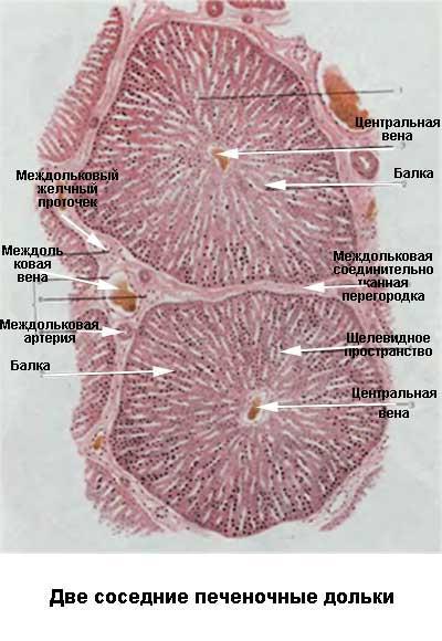
PD is the smallest morphological unit of the hepatic parenchyma. Visually has a prismatic shape. In its corners you can see the so-called portal, portal channels. There are five elements in them:
- Vein interlobular.
- Interlobular artery.
- Bile ducts in the hepatic lobule.
- Portal vein branch.
- A branch of the hepatic artery.
- Nerve fibers.
- A number of lymphatic vessels.
We will talk more about the structure of the lobule further.
The structure of the structural segment of the liver
The constituents of the lobule itself, in turn, are hepatocytes, specific polygonal liver cells. They are quite rather big sizes - 15-30 microns. A fifth of them are binuclear, 70% are mononuclear with a tetraploid set, the rest have a 4- or 8-fold diploid chromosome set.
Hepatocytes form hepatic platelets limited by sinusoidal hepatic capillaries. In the hepatic lobule, such plates have a thickness of one layer of hepatocytes. They are necessarily limited to endothelial cells and cells of the Kupffer hepatic sinusoids.
Examining the structure of the hepatic lobule, we see that these plates arise from a number of hepatocytes that limit the lobule from the side of the stroma, namely, the limiting plates. Having examined the latter on the anatomical atlas, we notice that they are speckled with a large number of holes. It is through them that the blood capillaries enter the lobule, while forming an already hepatic sinusoidal capillary network.
Liver plates and sinusoidal capillaries converge to a vector of the central vein passing through the organ.
Lobule blood supply: functional circulation
Blood supply to the liver lobule and the entire organ is entirely organized as follows.
Functional circulation (80% of the total proportion of the passing blood volume). The portal vein is divided into interlobar branches. Those, in turn, branch into interlobular, passing in the portal canals. Interlobular branches at strict intervals diverge into short perpendicular branches. They are called interlobular (entrance) venules. They cover the entire segment of the hepatic lobule.
From interlobular venules and veins, venous capillaries go to the surface of the lobule. It is with the help of them that blood passes through holes in the limiting plates into the sinusoidal capillaries of the liver. Further, it circulates between the liver plates and collects in the central vein.
From CV, blood is transferred to the submandibular vein, from where it enters the collective. In the end, it expires in the hepatic veins.
The role of the described functional circulation in the following:
- Delivery of absorbed nutrients from the digestive system, spleen, pancreas to the liver segments.
- Transformation and accumulation of metabolites.
- Neutralization and removal of toxic substances.
Lobule blood supply: nourishing circulation
The nutritional circulation of the hepatic lobule accounts for 20% of the total volume of blood passing through the segment.
The branches of the interlobar and hepatic arteries diverge into smaller branches - the interlobular arteries, whose path also lies through the portal canals. In turn, they are divided into arterial capillaries. The latter supply fresh, oxygenated blood to the portal canals, bile ducts, and organ stroma.
The next step is the collection of blood in the capillary web, which is formed by the entrance venules and interlobular veins. However, a small part of it in this case (mainly from the interlobular arteries) enters the sinusoidal capillaries. This helps to increase the oxygen content in the venous blood, which rotates in the hepatic sinuses.
Gate canal
The portal canal is a rounded or triangular space that can be seen in the corners of the liver lobule. VK is filled with connective loose tissue in which fibrocytes, fibroblasts, and vagus cells are located.
Through each channel pass:
- Bile duct.
- Interlobular vein and artery.
- Lymphatic vessels.
- Nerve fibers.
Let's talk about each of the presented units in detail.
Gateway blood supply
The blood supply to this part of the lobed parenchyma is represented by the interlobular artery and vein.
From the interlobular vein, capillary vessels extend, penetrating into the limiting plate, from where on to the hepatic lobule in the form of already sinusoids. Lateral vein branches located perpendicular to it - the entrance venules also turn into capillaries, becoming sinusoidal, with visible red blood cells.
The interlobular artery is of a muscular type, smaller in diameter than a vein. Capillaries supplying both the connective tissue of the portal canal and its contents also branch out from it. Part of the arterial branches is formed mainly in sinusoidal capillaries.
The capillaries from the arteries surround the bile duct, folding into the vascular peribiliary plexus.
Arterial and venous capillaries here have a similar structure. Hepatic sinusoids are actually sinusoidal capillaries. They pass between the plates of the liver so that their endothelium is separated from the plate only by a narrow Disse space - a perisinusoidal gap.
In the areas of bifurcation of the vessels of the hepatic sinusoids, specialized macrophages called Cooper cells are arranged in a chaotic manner. In the wide areas of the Diss cells, there are ITO cells, fat-containing or perisinusoidal.
Bile ducts
The bile ducts in the liver segments are always located between the bodies of hepatocytes and pass through the middle part of the liver plate.
The terminal bile ducts, distinguished by the fact that they are very short, are called Herring's canals. Lined with a small number of flat cells. It becomes visible Herring's channels only at the level of the bounding plate.
These terminal bile ducts exit already into the full bile ducts, which, passing through the portal canal, flow into the interlobular duct of bile. In the anatomical atlas, they are visible on the dissected liver plate as small holes.
Lymphatic and nervous system of the portal canal
Initial lymphocapillaries blindly begin inside the portal canal. Then they, having already separated from the restrictive plate by a narrow gap called the Mull space, are formed into lymphatic vessels. It should be noted that there are no interlobular ones among them.
Blood vessels accompany the nerve fibers of the adrenergic type, while innervating the portal canal itself. Then, passing into the hepatic lobule, an intralobular web is formed inside it. Cholinergic nerve fibers are also included in the lobule.
Lobule functions
The functions of the hepatic lobule are also the functions of the whole liver, since it is an integral segment of this large gland. The range of tasks of the body, as well as its components, is very wide. We will touch on the main functions that are most important for the body:
- Protection - activation of hepatic lymphocytes.
- The metabolism of active biological substances, the exchange of mineral elements.
- Participation in pigment exchange. It manifests itself in the capture of bilirubin and its excretion together with bile.
- Carbohydrate metabolism. Participation in the process involves the formation and subsequent oxidation of glucose, as well as the synthesis and breakdown of glycogen.
- Synthesis of bile, bile acids, triglycerides, phospholipids. All these elements are involved in both the digestive process and fat metabolism.
- The synthesis of a wide range of proteins necessary for the life of the whole organism - coagulation factors, albumin, etc.
- The most important is the cleansing, detoxification function. It is the liver - the main organ that cleanses the entire body of toxins. Through the portal vein, harmful, foreign substances, metabolic products enter the liver segments from the gastrointestinal tract. In this organ, they are further neutralized, after which they are excreted from the body.
The hepatic lobule is a component of the liver body. The body has a complex structure. The capillaries, lymphatic vessels, bile ducts and nerve endings pass through its portal canals. The lobule is based on special liver cells - hepatocytes, which have their own unique structure. The functions of both the entire liver and its lobules are similar.