Individual bones in the human body are connected to each other into a single skeleton by muscles, ligaments, and cartilage. They give it shape and strength, and in most cases mobility. Almost all bones in the human body are directly articulated.
Types of bone joint
There are 3 types of joints between the bones: fixed, semi-mobile and mobile. And the degree of mobility directly depends on the type and structure of the tissue located between the bones. It can bind them completely rigidly, provide limited flexibility of the connection or a wide range of movements:
- Fixed connection of bones has the property of being formed by their fusion, it is characterized by limited movement, its complete absence or slight mobility.
- Semi-movable joints exist between fixed and movable joints.
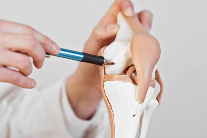
- Joints include mobile joints; they are responsible for the mechanics of movements in the body.
Fixed connection
The fixed junction of the bones has the human skull and pelvis. In them between the joints is a thin layer of a connecting seam or cartilage. Given the shape of the joint, there are jagged, scaly, and flat seams. All of them are the area of development of the bones of the skull and reveal a cushioning effect during movements.
Thanks to a fixed junction of bones has a skull, its bones protect the brain and sensory organs from external influences, and also form facial features. The only exceptions are the movable temporomandibular joints, which connect the lower jaw to the skull.
The structure of the bones of the skull
In the skull there are 2 important departments:
- One of them, the brain, is built from 8 bones that protect the brain and inner ear. All its bones are interconnected by layers of fibrous tissue - sutures.
- And the facial consists of 14 bones, where the nose, mouth and eyes are located. Bones of the facial section determine facial features of a person. The nasal cavity is divided into two symmetrical halves by a bone-cartilaginous septum. Bone outgrowths, called nasal concha, cause the flow of incoming air to warm and filter, leaving dust and germs on the mucosa. The paranasal sinuses open here .
Fixed joint of bones has a thin layer of fibrous tissue, which plays the role of glue. It is a dense fibrous mass that makes them motionless. Such a fixed junction of bones is called synarthrosis. Synarthrosis is found in the skull, for example, its sutures that have connective tissue between them. This is how the bones of the skull and teeth are combined with the holes of the jaws.
Semi-mobile connections
A person has 7 cervical vertebrae, of which two are superior: the atlas and axial. Atlas is an annular vertebra that carries the entire weight of a person’s skull. Its name, by the way, is dedicated to the mythical titan, holding the vault of heaven on his shoulders. It is thanks to the articulation of the atlas with the skull that a person can nod. And the axial vertebra, or epistrophy, forms a tooth from above, around which the atlas rotates with the skull.
The semi-mobile connection of the bones represents the cartilaginous tissue, but there is a shallow cavity in the thickness of the cartilage. Limited mobility of these compounds occurs due to cartilage plates and elastic ligaments.
Synchondrosis is a semi-mobile connection between the bones with a very low degree of mobility. Cartilage joints have limited mobility due to the elasticity of the cartilage connecting the bones. For example, a semi-mobile joint of bones has a pubic fusion closing the front of the pelvis and intervertebral discs. In the cartilage tissue, the cells are separated by an intercellular substance, which often forms a strong frame, allowing it to perform binding, supporting and protective functions.
Cartilage joints
There are several types of cartilage:
- Hyaline (vitreous) - the most common. It is very durable, smooth, bluish. It forms the skeleton of the embryo, and in adults - part of the ribs, airway support and articular surfaces of the articulated bones. Interestingly, the skeleton of the fetus consists mainly of such flexible cartilage, which gradually develops into bone tissue. The remains of the cartilage between the diaphysis and pineal gland become ossified by only 25 years. Moreover, not all skeletal cartilage is ossified. It remains on the articular surfaces of the bones, in the nose and auricles.
- Fibrous (fibrous) - whitish in color. It contains more collagen fibers and forms compounds with limited mobility, for example, connects the intervertebral discs. Such cartilaginous intervertebral discs may contract and twist slightly, but due to their large number provide flexibility of the spine.
- Elastic (mesh) - yellowish in color. It features a large number of elastin fibers, which gives it flexibility. Forms the skeleton of the auricle and part of the larynx.
Collagen is a very strong and flexible material that serves as the basis for most of the connective tissues that bind, divide and protect vital organs. These include bone, cartilage, fibrous tissue.
Fixed bone joint: examples
Speaking about motionless connection of bones, it is necessary to stop on synostoses, which are formed from syndesmosis and synchondrosis with age. In this case, the connective tissue or cartilage between some bones turns into bone.
So, the bones of the pelvic girdle are an example of which joints of the bones are motionless. They form something in the form of a bowl that protects and supports the organs of the lower abdominal cavity. This belt can withstand huge loads, the depression of the hip joint is much deeper than the shoulder, so it is much more stable.
For example, the tailbone consists of 3-5 rudimentary vertebrae, which grow together into a single triangular bone with age. Another example of synostosis is overgrown sutures or fontanel in an infant. These are physiological synostoses.
But with some diseases of the bone apparatus, ossification of not only cartilaginous tissue, but also of some joints can occur. These are pathological synostoses. Movement with such ossification, of course, are absent.