Magnetic resonance imaging is one of the most advanced and efficient methods for diagnosing diseases of the brain, especially of neurological origin. The MRI method consists in examining certain layers of the brain, as well as detecting abnormalities in its structure. A traditional X-ray does not show what the MRI of the head shows .
What is the installation
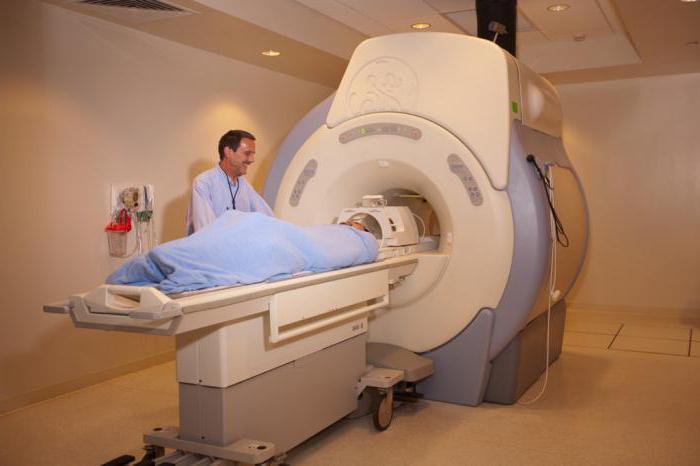
A typical tomography apparatus looks like a huge cylindrical tube surrounded by a magnet. The patient lies on a moving research table sliding inside the magnet. Certain tomographs (structures with a small tunnel) are produced in the order in which the magnet does not cover the patient’s table in full.
There is an MRI device that is open on the side. Such a system is especially good for inspecting fat people and those who are afraid of enclosed spaces. The current installed magnetic resonance tomographs make it possible to make excellent pictures with different examinations. However, if an outdated magnet is used in the installation of a simple model, the quality of visibility may decrease. There are studies that cannot be carried out on an open device.
Manifestations for performing tomography
Indications for MRI of the vessels of the head can be:
- chronic headaches;
- head injuries;
- frequent dizziness;
- seizures similar to epileptic;
- vascular health disorders;
- stroke;
- turbidity and loss of consciousness;
- analyzes confirming the presence of formations;
- increased or decreased pressure.
With the above signs, an MRI scan should be done sooner. A brain tumor, which can arise as a result of untimely treatment by a doctor, is far from a comic matter. If the tomography indicates the absence of complications in the head, then it is necessary to look for the cause in other places.
In what areas is MRI used
Magnetic resonance imaging is used to identify certain diseases and conditions, such as:
- detecting the origin of headaches;
- diseases of the inner ear and eyes;
- stroke;
- malfunctions and vascular pathologies of the brain (for example, aneurysms);
- malaise of the pituitary gland;
- injuries
- certain systematic diseases of the nervous system (multiple sclerosis);
- brain tumors.
How to prepare for the procedure
Special preparation is required only when examining the abdominal cavity and pelvic organs. Three days before the examination, you will need to follow a carbohydrate-free diet, and one day - take light food, also do not drink strong tea and coffee so that the doctor can determine what shows the MRI of the patient’s head. The patient is still not allowed to drink water and food 5 hours before the event.
Preparation for tomography under anesthesia
In this situation, there are also certain training rules, namely:
- be sure to consult a therapist;
- take the necessary tests;
- drinking is allowed no more than 1 cup 2 hours before the examination;
- food can be taken 9 hours before the study;
- if the patient wears contact lenses, then he must inform the anesthetist about this;
- Do not clean the skin with care products, in particular, do not apply cosmetics;
- leave in the next room all removable metal accessories (including jewelry, false jaws).
How is the survey
How to do brain MRI and how is it done? Now this will be discussed in more detail. Tomography can be performed both in the order of hospitalization of the patient, and on an outpatient basis. The radiologist assistant puts the patient on a movable table, his body position is fixed with additional rollers and belts, contributing to his calm position. Modules with wires that receive and send radio waves are located around the circumference of the body area under examination. With MRI of the brain, the installations are located near the head.
If there is a need to use a contrast agent during the examination, then the nurse can insert a catheter into the vein on the arm. A physiological saline bottle can be connected to the tube, which provides uniform flushing of the system. This eliminates clogging before the delivery of contrast material. At the end of all preparatory manipulations, the patient’s table is transferred into the magnet, where the MRI device itself is located. The radiologist and the minor medical staff during the examination leave the treatment room.
After the end of the study, the doctor sends the patient to the corridor to wait for the completion of the result of the obtained images, as an additional series of images may be needed.
The tomography procedure, as a rule, consists of the production of sequential images. Each of them takes several minutes. And the event itself usually lasts about 45 minutes.
In addition to brain MRI (only good reviews can be heard about it), magnetic resonance spectroscopy is also performed to evaluate the biochemical effects inside the cells. This inspection takes an additional 15 minutes.
Advantages of Magnetic Resonance Imaging
MRI has several advantages.
- Tomography is a non-invasive imaging technique that is not associated with ionizing radiation.
- MRI makes it possible to detect abnormalities that are invisible due to bone in other imaging methods.
- Magnetic resonance images of the brain, as well as other parts of the head, come out more detailed and expressive than with other systems. This side makes tomography a valuable tool in the initial diagnosis, detection of various diseases, as well as tumors.
- The contrast agent used in the examination in order to see what the head MRI shows shows less allergic effects than the iodine-based contrast material used for standard x-rays.
- Magnetic resonance imaging can determine the stroke at the very initial stage, showing the movement of microparticles of water in the tissues. This movement of water, called diffusion, is destroyed by most strokes.
- MRI helps doctors monitor the state of the structural parts of the brain.
- One version, called magnetic resonance angiography (MRA), provides a thorough visualization of blood vessels in the brain, often even without a contrast medium.
- MRI of the head, the price of which is today from 3,500 rubles. up to 5000 rubles., It is mainly an undoubted tool for determining brain tumors.
What are the risks?
- If anesthesia is used, there is a chance of excessive sedation. A technologist or nurse monitors vital functions to minimize this possibility.
- The MRI procedure is practically harmless for the average patient, if you follow the safety rules.
- There is a very small risk of allergic effects if contrast media is introduced. These reactions, as a rule, are little developed and are checked without any problems with the help of medications. And while the results of brain MRI are clearly visible.
- Currently, a side effect of tomography is nephrogenic systemic fibrosis. However, this does not happen often, and such a problem is revealed as a result of the introduction of a huge dosage of contrast medium and gadolinium in patients with very poor kidney function.
- Despite the fact that a powerful magnetic field is not harmful, implanted medical devices that contain metal can cause breakdown or create inconvenience during MRI.
Who can undergo the procedure
Magnetic resonance imaging does not harm a person if safety measures are followed. Patients with heart surgery or patients with certain medical settings can easily go through the procedure and find out what the MRI of the head and other organs shows. Valid devices:
- heart valve (with the exception of a cell or a metal ball);
- surgical sutures or clamps;
- hollow vein filters;
- artificial joints;
- shunt tubes for hydrocephalus;
- staples;
- disconnected drug pumps.
Contraindications
Under certain requirements, an MRI will only be harmful if there are:
- auditory implants;
- pacemaker;
- metal things in the eyes;
- excess weight more than 140 kg;
- severe lung disease (bronchopulmonary dysplasia, tracheomalacia);
- pregnancy;
- gastroesophageal reflux disease;
- transplanted insulin pumps (for the treatment of diabetes);
- cerebral clip of aneurysm (steel clip on a blood vessel in the head);
- claustrophobia;
- implanted rod for hardening the spine.
If the child has an MRI of the head (the price of such a procedure will depend on the location, but on average it will be from 2500 rubles), then you need to consult a tomography technologist in advance. Because it is very difficult for children to withstand a long period without movements. In such a situation, a sedative medicine is used - sleeping pills, sedatives or anesthesia. Also, parents, if necessary, are allowed to be with the child in the MRI room for the duration of the examination.