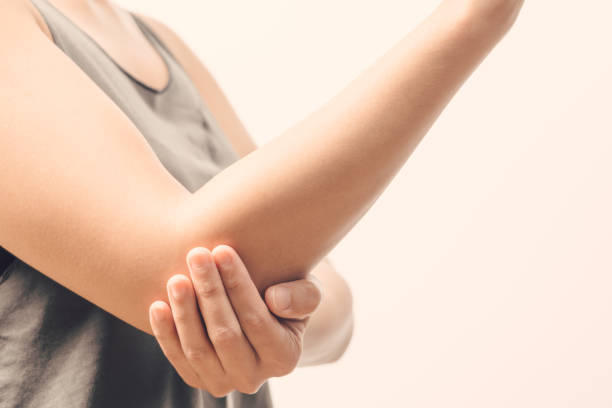
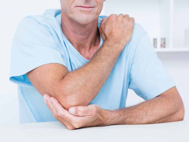
If we study the statistical information about fractures of the forearm, the radius (from the Latin radius) with almost the same structure and anatomy breaks much more often than the ulnar. This is due to the psychological feature of each person during the fall to put forward his hands, then the most severe blow will fall on that part of the surface on which the bone goes. Despite the fact that it is not a support to the body, like the lower extremities, its proper functioning affects the ability to move hands.
Most often, it is the head of the radial bone that suffers.
The location of the radius
Radius in the forearm is located adjacent to the ulna. That is why they are dependent and interconnected with each other. When the palm is turned back with the raised hand, they are both parallel, however, when the palm is turned in the opposite direction, the bones cross. Partially, the beam rotates near the ulnar, which ensures pronation (repeated ability) and supination (rotational capability). In addition, the location of the radius can be determined on the thumb of the hand.
Radial bone anatomy
The radius includes the diaphysis (long body) and the two ends - the proximal and distal. The distal pineal gland is more powerful, on it lies the surface of the wrist joint, as well as the styloid process, which connects to the hand. The structure of the proximal end of the radius is as follows: the articular circumference (the beam connects with the humerus with its help) and the head. Under the head of the radius is the neck, then tuberosity, to which the brachial biceps muscle is attached. Its development is due to the development of ossification points. Three types of faces are distinguished: the back (with a rounded edge), the front (also rounded) and the lateral (the face goes to the ulnar, with a pointed edge).
Definition and Functions
The head of the radial bone is the uppermost portion of the bone, it is one of two bones of the forearm. The main function is to provide rotational movements in the forearm (palm down - palm up). Its form is determined entirely and completely by its function.
Fracture
A fracture of the radial head means a violation of the bone structure, which is located in the elbow joint. This is a lesion inside the joint.
The following types of disease according to the specifics of trauma:
- fracture of the radial head without displacement;
- with offset;
- comminuted;
- regional.
Fractures according to the degree of openness are divided into closed and open.
Treatment methods
Conservative treatment can be dispensed with depending on the degree to which fragments are displaced (if the displacement is small) - six to eight weeks in a plaster cast.
Then you need a rehabilitation course - special exercises to help develop the joint.
If there are fractures with a strong displacement of the radial head, then there are two methods of surgical treatment - removal or fixation of fragments of this head (osteosynthesis). Surgical therapy is carried out through a five-centimeter incision along the outer surface of the forearm. The doctor decides on the choice of a particular method, based on a large number of factors - the occupation of the person, the degree of displacement of fragments, etc.
In most cases, the removal of the head does not affect the functioning of the forearm very much - a person in everyday life almost does not feel any inconvenience.
With a significant displacement of fragments, a fracture of the radial head of the elbow joint can be fixed by means of small knitting needles or screws (osteosynthesis).
When the fracture is too complex, with a large number of small fragments that are almost impossible to connect, the radius head is removed. In some cases, a prosthesis is placed instead, but according to the latest information, it is not always necessary to replace it with the forearm to restore normal functioning. Postoperative therapy consists in the use of calcium preparations, painkillers, sometimes topical preparations to reduce swelling. After the operation, the guard mode is that the hand is worn for about three weeks on a scarf, sometimes a plaster cast is used .
How to develop a limb
After three weeks, the patient needs to actively develop the arm. For this, physiotherapy exercises can be prescribed, which increases the range of movements, physiotherapy courses (for example, magnetotherapy, phonophoresis with hydrocortisone and cryotherapy) to improve the condition of soft tissues.
Next, consider the subluxation of the radial head.
Dislocations and subluxations
Isolated dislocations and subluxations of the upper extremities are most often observed in childhood. The trauma is sometimes so small that parents do not pay attention to implicit symptoms, and then a subluxation, including the radial head, develops into a chronic trauma. You need to be very careful about the complaints of the child, if any. So, in preschool children, a dislocation of the radial head is one of the most common injuries that occur during a fall. Isolated complete dislocation in medical practice is observed in children under the age of five. Dislocations of the radial head are divided into acquired and congenital.
Congenital patients are less frequent and most often easily restored if they were previously recognized. Sometimes it turns out to cure subluxation without the use of significant medical manipulations. When leaving the disease unattended, we are talking about a long-standing dislocation. His danger is that he gives the joint area vulnerability, limits the motor functions of the joint.
Adult patients are more likely to have dislocations of the radial head, and in young children, subluxations. The majority of injuries in this case are pronational dislocations that the baby receives when stretched. Front injuries in the elbow joint are noted when falling on the hands. The specifics of a typical dislocation of the head of the radial joint radial bone in patients from one to five years are damage to the ligaments, in girls with such injuries are twice as likely.
Features of diagnosis and treatment
Reduction in some cases is carried out without preliminary diagnosis by instrumental means. An x-ray is required for an unspecified type of injury or suspected possible fracture. Congenital dislocations in obstetrics are frequent, if the inflammation progresses in the fetus or there is intoxication of the body, the help of a toxicologist is needed. The newborn can be diagnosed with a “subluxation of the radial head” by palpation. Ultrasound is sometimes prescribed. Often injuries require differential diagnosis. In children after three years of injury, they have similar symptoms. In this case, radiography is mandatory.
The damaged area is clearly visible in the image, and the X-ray diffraction pattern also shows chips and fissures of the bone structures. Treatment involves such manipulations: the use of painkillers - the pain experienced by a child after an injury is suppressed by non-narcotic analgesics. Ibuprofen preparations are often given, which are also characterized by anti-inflammatory effects. A closed reposition is carried out - the shoulder joint is fixed before interspersing.
The reposition is done in a sitting position, a healthy arm is extended along the body. In this case, a smooth movement is carried out in the elbow region of the injured limb, palm down, to the absolute supination. The head of the radius after implantation falls into place, a click is heard. Another way is immobilization, specific treatment is not required, an overlay for three days of a scarf dressing is enough. With a complicated injury, they use a plaster cast, they wear it for up to three weeks.
The treatment of dislocation of the radial head is selected based on the medical history. Complex manipulations in early childhood are not needed. In infants, the joint can be repaired the first time. To avoid a new dislocation of the radial head in childhood, use bandages and orthoses, the wearing period of which is determined individually.
Operative therapy
If complications arise after an injury and treatment, consult a doctor. With a dislocation of the radial head with a concomitant fracture of the elbow bone, healing is more complicated. Closed reposition can sometimes not be done due to the presence of bone fragments and atypical displacement of the joints. The operation is also required in case of rupture of the ulnar process, violation of nerve fibers and blood vessels, improper fusion of an old injury. With a constant displacement of the head, you need to fix it with knitting needles. The same procedure is required for a complicated fracture in the elbow. After surgical treatment, the recovery period is longer.