Ancient healers could not even imagine that in the future it would be possible to examine the internal organs of a person and at the same time not to make incisions on the body. Currently, such a survey has become a reality. Medical science is constantly evolving, due to which it is possible to timely identify various pathological conditions and provide the necessary assistance to patients. Endoscopic examinations allow you to assess the condition of the tissues of the hollow organs from the inside. There are several varieties of such diagnostics that will be discussed in this article.
What is endoscopy?
In medical practice, the term "endoscopy" means an examination of internal organs with a cavity using lighting devices. To perform this procedure, an endoscope is used - rigid or flexible tubes of small diameter. In the first case, the basis of the device is an optical fiber system. On one side is a light bulb, and on the other, an eyepiece that allows you to adjust the image size. Flexible endoscopes allow you to explore the most inaccessible places. A clear picture is transmitted through the fiber bundle despite the bending of the system. A new step in the development of this diagnostic area is capsular endoscopy.
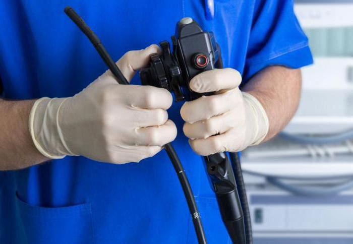
With the help of flexible endoscopes, you can not only diagnose, but also take tissue samples (aspiration biopsy) for a more detailed study of the pathological process. Endoscopic studies can determine the nature of the disease, track the dynamics of treatment. A unique device allows you to evaluate the condition of almost any organ. The procedure itself is carried out exclusively in medical institutions by specially trained personnel.
Method Advantages
The main advantage of diagnosis with an endoscope is the ability to see the status of internal organs without surgical intervention. The procedure is painless for the patient. The only thing he can feel is discomfort. In the process of examination, the person is conscious.
The diagnostic method is sometimes used for operations. In this case, a small skin incision is made, through which a tube with a lighting device will be introduced. Such manipulation is necessary when removing benign neoplasms on the internal organs, while removing foreign bodies. Endoscopic research methods can be used to administer drugs.
Endoscopy Applications
The appearance of endoscopy made it possible to examine almost all organs. The diagnostic method is used in the following areas of medicine:
- gynecology (coloscopy, hysteroscopy);
- neurology and neurosurgery (ventriculoscopy);
- pulmonology (bronchoscopy);
- otolaryngology (otoscopy, pharyngolaryngoscopy);
- gastroenterology (gastroscopy, colonoscopy, esophagogastroduodenoscopy, laparoscopy);
- cardiology (cardioscopy);
- Urology (cystoscopy, ureteroscopy).
Recently, endoscopy is also used to diagnose knee joints. In the process of diagnosis (arthroscopy), a special device is introduced to the patient - an arthroscope, which allows the specialist to assess the condition of the joint and perform the procedure with minimal surgical intervention. Conducting endoscopic examinations also allows you to recognize an ailment at an early stage, therefore, they are often prescribed for prevention in patients at risk.
Indications for bowel examination
The only way to see the condition of the intestine is to have an endoscopy. In medical terminology, endoscopic studies of this kind are called esophagogastroduodenoscopy, colonoscopy, and sigmoidoscopy. Indications for the diagnosis of the esophagus, stomach, colon and small intestine, rectum are the following pathological conditions:
- Peptic ulcer.
- Suspicion of bleeding.
- Oncological diseases.
- Gastritis.
- Paraproctitis.
- Violations of the stool.
- Hemorrhoids (chronic).
- Discharge of blood, mucus from the anus.
Depending on the preliminary diagnosis, the specialist will choose the most suitable option for endoscopic examination.
Intestinal colonoscopy
One type of endoscopic examination is colonoscopy. The method allows you to diagnose the large intestine using a flexible colonoscope device, consisting of an eyepiece, a light source, a tube through which air is supplied and special forceps for collecting material. The device allows you to see a fairly high-quality image displayed on the screen, the state of the mucous membrane of the colon. The length of the tube used for this type of diagnosis is 1.5 meters.
The procedure is quite simple. The patient is offered to lie on his left side and pull the legs bent at the knees to the chest. Then the doctor carefully inserts a colonoscope into the rectum. Anus can be pre-lubricated with anesthetic gel. The tube is gradually advanced inward, examining the walls of the intestine. For a clearer image, air is constantly supplied during the diagnostic process. The procedure takes no more than 10 minutes.
Do I need training?
Of course, to obtain an accurate picture of the condition of the large intestine, the patient should be prepared for colonoscopy. Preparation for endoscopic examination consists primarily in following a diet. Exclude from the daily menu products that contribute to the delay of feces and increased gas formation should be done at least one week before the expected date of the diagnosis.
On the day of the examination, you need to refrain from the morning meal. Allowed only to drink liquid. Before the procedure itself, experts recommend cleaning the rectum with an enema or using laxatives.
An endoscopic examination of the intestine - colonoscopy - is a painless procedure and therefore you should not be afraid of it. The patient may feel only slight discomfort. In some cases, the manipulation is carried out under anesthesia, but most often they are limited to sedatives and painkillers.
Capsule endoscopy
A relatively new direction in the diagnosis of diseases of the gastrointestinal tract is capsular endoscopy. The method appeared only in 2001. The endoscope used for research resembles a drug capsule, which greatly facilitates the process of introducing the device. Such a tablet just needs to be washed down with water. The device is activated immediately after opening the individual packaging. Passing through the organs of the digestive tract, the capsule takes many pictures, which in the future will help to make a diagnosis.
The advantages of this method are obvious - the patient does not need to swallow the hose or worry about having a colonoscopy. The capsule enters the most remote parts of the intestine, where there is no access to the usual endoscope. On the other hand, this method does not allow you to take material for a biopsy, remove polyps. Therefore, doctors still prefer to comprehensively use capsular and traditional endoscopy of the digestive tract.
Esophagoscopy
An endoscopic examination of the esophagus is performed to diagnose various pathologies. Esophagoscopy is most often combined with an examination of the stomach and duodenum. This allows you to get a more complete picture of the state of the digestive tract. The method allows to identify ulcers, hemorrhages, inflammatory processes, polyps on the mucous membrane. Taking material for a biopsy allows you to establish the etiology of the disease. Inspection is carried out by both a flexible and a rigid device.
Indications for examination are structural abnormalities, gastroesophageal reflux, chemical burns of the mucosa, the need for a biopsy, the presence of a foreign body, and inflammatory processes.
Endoscopic Ultrasound
To diagnose the walls of the digestive tract, an endoscopy method using ultrasound can be used. The latter allows you to get an image of organs thanks to sound waves. This method is most often used to detect benign neoplasms, tumors, stones in the bile ducts, and inflammation of the pancreas. Ultrasound endoscopic examinations make it possible to evaluate the mucous membrane of the entire digestive system.
An endoscope is introduced to the patient through the larynx first into the esophagus, gradually moving it into the stomach and duodenum. Previously, the larynx is treated with an analgesic spray to stop unpleasant sensations. Ultrasound may be needed to take tissue samples.
The consequences of the procedure
Endoscopic research methods in most cases do not cause serious disturbances in the body. With the correct procedure, the patient can return to a normal lifestyle within a few hours and not feel any unpleasant sensations. However, there are still situations when, after a diagnosis, a person is forced to seek medical help. Most often, damage to the walls of organs when passing through an endoscope is recorded. This can be determined by the pain syndrome, which does not pass for a long time, the presence of blood in the feces.
An allergic reaction to the analgesic used in the study may occur. In this case, the use of antihistamines is indicated. Arrhythmia after the procedure often develops in patients with cardiovascular pathologies.
Proper preparation of the patient for endoscopic examinations will avoid many undesirable consequences. Diagnosis itself should be carried out in a hospital or clinic. Previously, the doctor must exclude all contraindications for this kind of examination.