If you suspect the development of certain diseases (often infectious), the patient takes an analysis of the cerebrospinal fluid, which is called cerebrospinal fluid. The procedure is safe for humans. However, it has certain features and side effects. To draw conclusions about the features of conducting such a study, the procedure and analysis norms will be discussed in detail below.
Cerebrospinal fluid function
Before you consider how to take the analysis of cerebrospinal fluid, you should find out what function it performs in the body. Liquor is also called cerebrospinal fluid. This is a biological element that is constantly located and circulates in the ways allotted for it. It is concentrated in the subarachnoid membranes of the brain, spinal cord. Also, cerebrospinal fluid is present in the ventricles of the brain.
Cerebrospinal fluid performs important functions for the human body. It provides a balance of the components of the internal environment of the two most important parts of the body - the brain and spinal cord. Liquor protects them from shock by absorbing mechanical shocks. With the help of it, the saturation of neurons (brain cells) with the necessary nutrients, oxygen. Also, the liquid removes carbon dioxide, toxins, and other substances spent during the metabolism from them.
Cerebrospinal fluid supports the optimal chemical composition of the internal environment, as well as the pressure inside the skull. It contains white blood cells that do not allow infections to develop inside the brain. The performance of these functions becomes possible only due to the constant flow of fluid in the paths. Liquor is constantly updated.
Analysis of cerebrospinal fluid allows you to determine the development of various pathologies. If you define them at an early stage, the treatment will be much faster and easier. It is worth noting that the amount of water that a person drinks per day affects the rate of cerebrospinal fluid composition. For the body to function normally, it needs 1.5-2.5 liters of water per day. In this case, the correct pressure is maintained inside the brain. Otherwise, the person feels unwell.
Normal performance
There are certain standards for the analysis of cerebrospinal fluid. In a healthy person, indicators should be within certain limits. If the cerebrospinal fluid does not meet the established standards, the doctor can diagnose a certain pathology. So, the cerebrospinal fluid should be transparent and colorless, visually similar to clean water. After examining the composition in appearance, proceed directly to the analysis of cerebrospinal fluid. The protein norm in it is up to 0.45 g / l. Cell composition is also evaluated. In 1 μl should be 1-2 lymphocytes. Glucose should be in the liquid from 30 to 60%. This indicator depends on the nutritional characteristics of the patient. To correctly examine this indicator, it is compared with the data of a blood test. The pressure in the system should be 100-150 cm of water.
In addition to microscopy, in the analysis of cerebrospinal fluid, its amount is examined. It should vary between 130-160 ml. This indicator depends on the physiology of the body.
90% of cerebrospinal fluid consists of water. Proteins, amino acids, glucose and lipids are dissolved in it. Also in the liquid is ammonia, traces of concentrates of nitrogen compounds and urea. Lactic acid is present in the cerebrospinal fluid, as well as the remains of cells and their individual fragments.
The density of the liquid is from 1003 to 1007 g / l. Also during the analysis, the reaction of the medium is determined. Normally, the pH is 7.37-7.88 units. The composition of the cerebrospinal fluid is alkaline. However, the indicator of environmental characteristics should not go beyond the established framework.
It is worth noting that the pressure standards may differ if the patient is in a sitting or lying position at the time of collection of the biological material. This phenomenon is due to the redistribution of body weight, which presses on the cerebrospinal fluid in different positions.
Cytosis in the analysis of cerebrospinal fluid can be from 1 to 10 μl. This indicator characterizes the number of cells in the fluid. They constantly enter the cerebrospinal fluid from tissues and blood. This is considered normal.
Indications for the study
A general analysis of cerebrospinal fluid is performed if a number of pathologies are suspected. After examination, the doctor may prescribe a similar procedure if the patient is suspected of having a tumor. The neoplasm can be located in different parts of the body. Analysis can confirm or deny its presence.
For traumatic brain injuries, a similar study is also required. If you suspect a heart attack or a stroke of the brain or the diseases that accompany them, the doctor may prescribe a similar procedure. One of the groups of indications is an infection in the lining of the brain. Therefore, an analysis of cerebrospinal fluid is almost always prescribed for meningitis, meningoencephalitis, etc.
The indication for the examination may be the presence of an intervertebral hernia, epilepsy, or brain hematoma. In the presence of such diseases, the analysis can detect the presence of pathology.
The collection of biological material is carried out by taking a puncture. The procedure can be carried out both for diagnostic and therapeutic purposes. Sometimes in the process of such a puncture an antibiotic is introduced into the body. It is worth noting that this procedure is completely safe. It does not lead to disturbances in the spine. Therefore, you should not be afraid that complications will arise after taking the cerebrospinal fluid. There is a certain technique for taking biological material.
In specialized clinics, on the basis of the examination, the doctor will be able to diagnose a number of diseases dangerous to human health and life. By comparing the indicators with the standards, you can determine the deviation. Next, its cause is established. This allows us to draw conclusions about the processes occurring in the patient's body.
How is the analysis carried out?
Many patients are interested in how cerebrospinal fluid analysis is done. This procedure is special. For its implementation, a doctor of appropriate qualification makes a lumbar puncture. A special needle is inserted into the tissue. In some cases, the patient is shown atlanto-occipital puncture.
The doctor drops the first drop on a napkin. This avoids ingestion of track blood. Her presence can significantly affect the result. Considering how to analyze the cerebrospinal fluid, it is worth noting that at the slightest suspicion of getting the test blood in the tube, the puncture is redone. Each time a new needle is used.
Due to some circumstances, it is impossible to take a puncture from some patients due to ingestion of blood in the material. If three attempts were unsuccessful, a fourth puncture is not performed. This can lead to the development of various complications.
Liquor is not collected in glass tubes. In this case, there is a chance that the white blood cells will adhere to the glass.
To take the required amount of fluid, a puncture is made in the lumbar region. It is safe to take a puncture here. Penetration of the needle into the membrane of the spinal cord does not harm a person. Here the nerve fibers move freely in the cerebrospinal fluid. It is impossible to pierce them with a needle. However, after a puncture, a person feels constant discomfort in the lumbar region. Headaches may also occur. Unpleasant symptoms disappear on their own in a couple of days.
The speed at which cerebrospinal fluid analysis results are obtained depends on the policy of the clinic in which the examination is conducted. Material is delivered to the laboratory no later than an hour after the puncture. Usually the patient receives the test result the next day.
Analysis Kit
To perform such an analysis, a set of reagents is used to analyze cerebrospinal fluid. It includes a number of components that interact with biological material. The cost of such sets varies from 1200 to 1500 rubles. By default, it can be used to determine the following:
- cytosis;
- quantity and quality of indicators of protein;
- a qualitative indicator of globulin.
To prevent cell cytosis for several hours, Samson's reagent is used. It is part of almost every set for cerebrospinal fluid analysis. The reagent contains acetic acid. It dissolves red blood cells. The reagent also contains fuchsin, which stains the nuclei of cells in red. In this case, it is much easier for a laboratory assistant to consider their amount in biological material. Also, without problems it turns out to perform cell differentiation.
Qualitative protein analysis is carried out using the Pandy reaction. The kit for clinical analysis of cerebrospinal fluid contains phenol. It reacts with protein. As a result, the liquid becomes cloudy. The more intense this process, the correspondingly more specific protein is in the cerebrospinal fluid. In a similar way, its quantity in the composition is also determined. Only in this case sulfosalicylic acid and sodium sulfate are used. The more turbid the composition, the more protein it contains.
To check the composition of globulins, the None-Apelt reaction is used. Biological substances react with ammonium sulfate. When using such sets, it turns out to determine how certain processes in the body proceed, whether there is any pathology. Decryption is carried out by an experienced doctor of relevant qualifications.
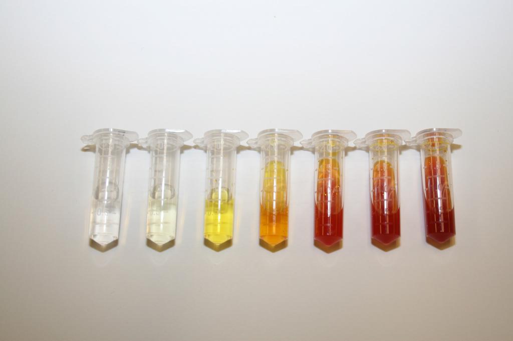
Fluid color
It is worth noting that the interpretation of the analysis of cerebrospinal fluid is carried out comprehensively. The indicators obtained during the study of blood, urine, as well as some instrumental procedures are compared. Patient complaints are also considered. One of the important indicators is the color of the cerebrospinal fluid. If the liquid is no longer transparent, it determines the increased viscosity, this indicates the development of the disease. According to what color the liquid is painted, we can talk about the development of certain pathologies:
- Red. In the subarachnoid space, hemorrhage is determined. High blood pressure is determined here. This condition indicates a pre-stroke condition.
- Light green. The liquid may also have a yellowish tint. This color indicates the development of meningitis or brain abscess. A similar situation occurs with complications of an inflammatory nature.
- Opalescent or scattering. Talks about the development of the pathological process. It develops in the shells of the brain. May also be present with bacterial meningitis.
- Yellow. It is called xanthochromic. The hue speaks of a brain hematoma or the possible development of oncology in this department.
If the liquid is cloudy, this indicates a large content of cells in it. Including it can be bacteria. A serious inflammatory process develops in the body. An increased density of cerebrospinal fluid indicates the presence of a head injury or inflammation. Too low a density is also a pathology. This condition is called hydrocephalus.
Cytosis, protein concentration
In the course of deciphering the analysis of cerebrospinal fluid, an indicator such as cytosis is necessarily investigated. An increase in the concentration of cells in biological material should not exceed certain norms. If the cytosis is increased, exceeds the permissible value, this may indicate the following:
- complications of stroke or cerebral infarction;
- allergy;
- the appearance of oncological neoplasms;
- meningitis;
- organic lesions of the meninges.
Also, the level of protein in the analysis is necessarily controlled. Its increased concentration indicates the appearance of serious pathologies. For example, it can be meningitis, benign or malignant neoplasms, hernia (protrusion) of intervertebral discs, encephalitis. Also, a similar situation can talk about squeezing neurons located in the spinal column.
Reducing the amount of protein in the cerebrospinal fluid is not a pathology. Fluctuations of this indicator in the negative direction are a physiological state. This cannot be regarded as a symptom of the disease.
Protein enters the cerebrospinal fluid from blood plasma. With its increase, the blood-brain barrier becomes permeable. Through it, protein enters the cerebrospinal fluid. This indicates the development of serious pathologies in the body. To make a correct diagnosis, an analysis of the protein content in the blood serum is carried out. Based on the information received, an albumin index is obtained. For this, the indicator of protein in cerebrospinal fluid is divided into a similar value in blood plasma.
Next, the degree of damage to the blood-brain barrier is assessed. If the index is less than 9, no violations were detected. If the indicator is in the range from 9 to 14 units, the lesion is regarded as moderate. Noticeable disorders are diagnosed in the presence of an albumin index of 15-31 units. Severe damage is determined in the range of 31-100. Over 101 units, impaired barrier function is complete.
To determine the amount of protein, the biological material is mixed with sulfosalicylic acid, sodium sulfate. As a result, the liquid becomes cloudy. The intensity of this process is determined photometrically. For this, special equipment is used. The result is evaluated at a wavelength of 400-480 nm.
Glucose and Chlorides
During the clinical analysis of cerebrospinal fluid, a glucose value is also determined. A negative phenomenon is considered both an excess and a decrease in sugar in the cerebrospinal fluid. If the norm is exceeded, we can talk about the development of various diseases. This can be epilepsy, concussion, cancer. In addition, an increase in glucose may indicate the development of diabetes of the second or first type.
A low sugar level in the cerebrospinal fluid indicates the development of an inflammatory process. Including it can have a tubercular nature. Similar symptoms are characterized by meningitis.
The analysis also determines the concentration of chlorides. Raising or lowering this indicator is unacceptable. If the chloride concentration in the biological material is exceeded, an additional examination is required. A similar situation may indicate the development of renal or heart failure, as well as cancer.
If the concentration of chlorides is reduced, this may indicate the development of meningitis. Also, a similar situation is observed with the appearance of a tumor. In this case, a set of indicators is necessarily investigated. A doctor cannot make a diagnosis solely on the basis of a deviation of one indicator. A comprehensive examination allows you to get the right result.
Microscopy
During the analysis of cerebrospinal fluid, they can count the number of cells and create a cytogram in smears. To do this, they are stained according to Noht or Romanovsky-Giemsa using azure-eosin. However, in addition to the number, the composition of the cells is also studied. For this, microscopy of biological material is performed.
In a normal state, only monocytes and lymphocytes enter the cerebrospinal fluid. However, due to various reasons, diseases, other cells may also be included. It should be noted that normally up to 10 lymphocytes are contained in the cerebrospinal fluid. Their number increases with the development of tumors in the central nervous system. Also, their level rises in the presence of an inflammatory process in the membranes of the brain.
Other cells
If blood plasma cells are determined in biological material, this indicates the development of an inflammatory process in the brain for a long time with encephalitis, meningitis, as well as a number of other similar diseases. A similar situation is observed in the postoperative period.
If tissue monocytes are present in the cerebrospinal fluid, this also indicates the development of a chronic inflammatory process in the central nervous system. Allowed single inclusion of these cells in the cerebrospinal fluid. If there are a lot of them, this indicates an active tissue reaction during wound healing.
Macrophages should also not be found in cerebrospinal fluid. They appear in the cerebrospinal fluid only after bleeding or inflammation. It is considered normal if such cells are in the biological material collected for the study in the postoperative process. .
. , . , , .
Eosinophils are present in the analysis in the presence of subarachnoid hemorrhage, brain tumors, and meningitis. Very rarely, epithelial cells are observed in the collected material. This is a sign of the development of a tumor or an inflammatory process.
Having considered the features of conducting and interpreting the results of the analysis of cerebrospinal fluid, we can expand our knowledge of this procedure.