Man is the most mysterious and studied organism on planet Earth. Each of its organs has its own task and continuously performs its functions: the heart pumps blood throughout the body, the lungs provide breathing, the esophagus and stomach are responsible for restocking, and the brain processes all the information. Consider the function of the organs of the chest cavity in the human body.
Chest cavity
The chest cavity is the space in the body that is inside the chest. The chest and abdominal cavities separate the internal organs that are located in them from the skeleton and muscles of the body, allowing these organs to move smoothly inside relative to the walls of the body. Organs located in the chest cavity: heart, blood vessels and nerves, trachea, bronchi and lungs; the esophagus passes from the chest cavity into the abdominal cavity through an opening in the diaphragm. In the abdominal cavity are the stomach and intestines, liver, kidneys, spleen, pancreas, numerous vessels and nerves.
The photo shows where and what organs of the chest cavity are located. The heart, trachea, esophagus, thymus, large vessels and nerves are located in the space between the lungs - in the so-called mediastinum. A domed diaphragm attached to the lower ribs, back of the sternum and lumbar vertebrae forms a barrier between the organs of the human chest and abdominal cavity.
Heart
The most working muscle of the human body is the heart or myocardium. The heart measuredly, with a certain rhythm, without stopping, distills the blood - approximately 7200 liters daily. Various parts of the myocardium simultaneously contract and relax at a frequency of about 70 times per minute. With intense physical work, the load on the myocardium can increase threefold. Heart contractions start automatically - a natural pacemaker located in its sinoatrial node.
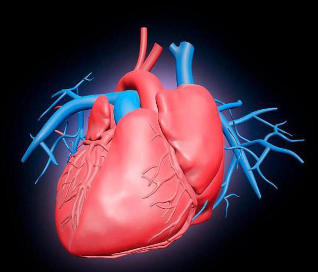
Myocardium works automatically and is not subject to consciousness. It is formed by many short fibers - cardiomyocytes, interconnected into a single system. Her work is coordinated by a system of conducting muscle fibers of two nodes, in one of which is the center of rhythmic self-excitation - a pacemaker. It sets the rhythm of contractions, which can change under the influence of nerve and hormonal signals from other parts of the body. For example, with a heavy load, the heart beats faster, sending more blood to the muscles per unit time. Thanks to its performance, about 250 million liters of blood are passed through the body over 70 years of life.
Trachea
This is the first of the organs of the human chest cavity. This organ is intended for the passage of air into the lungs and is placed in front of the esophagus. The trachea begins at the height of the sixth cervical vertebra from the cartilage of the larynx and branches on the bronchi at the height of the first thoracic vertebra.
A trachea is a tube having a length of 10-12 cm and a width of 2 cm, consisting of two dozen cartilages in the shape of a horseshoe. These cartilage rings are held in front and side by ligaments. The gap of each horseshoe-shaped ring is filled with connective tissue and smooth muscle fibers. The esophagus is located immediately behind the trachea. Inside the surface of this organ is covered with a mucous membrane. Trachea, being divided, forms the following organs of the human chest cavity: the right and left main bronchi, descending into the roots of the lungs.
Bronchial tree
Branching in the form of a tree contains the main bronchi - right and left, partial bronchi, zonal, segmental and subsegmental, small and terminal bronchioles, followed by the respiratory departments of the lungs. The structure of the bronchi differs throughout the bronchial tree. The right bronchus is wider and placed steeper downwards than the left bronchus. Above the left main bronchus is the aortic arch, and under it and in front of it is the pulmonary trunk of the aorta, which is divided into two pulmonary arteries.
The structure of the bronchi
The main bronchi disperse, creating 5 lobar bronchi. 10 segmental bronchi go further from them, each time decreasing in diameter. The smallest branches of the bronchial tree are bronchioles with a diameter of less than 1 mm. Unlike the trachea and bronchi, bronchioles do not contain cartilage. They consist of many smooth muscle fibers, and their lumen remains open due to the tension of the elastic fibers.
The main bronchi are located perpendicularly and rush to the gates of the corresponding lungs. At the same time, the left bronchus is almost two times longer than the right, has the number of cartilaginous rings 3-4 more than the right bronchus, and seems to be a continuation of the trachea. The mucous membrane of these organs of the chest cavity is similar in structure to the mucous membrane of the trachea.
The bronchi are responsible for the passage of air from the trachea to the alveoli and back, as well as the purification of air from foreign impurities and their removal from the body. Large particles leave the bronchi during coughing. And small particles of dust or bacteria that have entered the respiratory organs of the chest cavity are excreted by the movements of the cilia of epithelial cells, which advance the bronchial secretion towards the trachea.
Lungs
In the chest cavity are organs that everyone calls them lungs. This is the main paired respiratory organ, which occupies most of the chest. Separate the location of the right and left lung. By their shape, they resemble cut cones, with the apex directed towards the neck, and a concave base to the diaphragm.
The top of the lung is 3-4 cm above the first rib. The outer surface is adjacent to the ribs. The bronchi, pulmonary artery, pulmonary veins, bronchial vessels and nerves lead into the lungs. The place of penetration of these organs is called the gate of the lung. The right lung has a greater width, but shorter than the left. The left lung in the lower front has a niche under the heart. A substantial amount of connective tissue is present in the lung. It has a very high elasticity and helps the work of the contracted lung forces, which are necessary with each inhalation and exhalation.
Lung volume
At rest, the volume of inhaled and exhaled air averages about 0.5 liters. The vital capacity of the lungs, that is, the volume at the deepest exhalation after the deepest breath, is in the range from 3.5 to 4.5 liters. For an adult, the rate of air consumption is approximately 8 liters per minute.
Diaphragm
The respiratory muscles rhythmically increase and decrease the volume of the lungs, changing the size of the chest cavity. The main work in this case is made by the diaphragm. When contracting, it flattenes and falls, increasing the size of the chest cavity. The pressure in it drops, the lungs expand and draw in air. The lifting of the ribs by the external intercostal muscles also contributes to this. With deep and accelerated breathing, auxiliary muscles participate, including the pectoral and abdominal ones.
The mucous membrane of these organs of the chest cavity consists of the epithelium, and that, in turn, consists of many goblet cells. In the epithelium of the branches of the bronchial tree there are many endocrine cells that control the blood supply to the lungs and maintain the tone of the bronchial muscles.
To summarize all of the above, it is necessary to note that the organs of the human chest cavity are the basis of his life. It is impossible to live without a heart or lungs, and a violation of their work leads to serious diseases. But the human body is a perfect mechanism, you only need to listen to its signals and not harm, but help Mother Nature in its treatment and recovery.