If there is a system in the body, then there is something that fills it. The activity of structure branches depends on the quality of the content. This position can be fully attributed to the work of the circulatory and lymphatic systems of a person. The healthy content of these structures is an integral factor in the stable functioning of the whole organism. Next, we will examine in more detail what significance the blood and lymph vessels have. Let's start with the latter.
General information
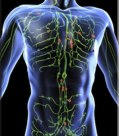
Human lymphatic vessels are represented by different structures that perform certain functions. So, allocate:
- Capillaries.
- Large trunks (thoracic and right ducts).
- Extra - and intraorgan vessels.
Also, the structures are muscle and muscular type. The flow rate and pressure (hemodynamic conditions) are close to those that take place in the venous channel. If we talk about the structure of the lymphatic vessels, it is necessary to note the well-developed outer shell. Due to the inner coating, valves are formed.
Capillary
This lymphatic vessel has a fairly permeable wall. The capillary is able to absorb suspensions and colloidal solutions. The channels form networks that represent the beginning of the lymphatic system. Connecting, capillaries form larger channels. Each formed lymphatic vessel passes to the subclavian veins through the neck and sternum.
Moving content in channels
The movement of lymph through the lymphatic vessels is carried out along the cervical duct into the venous bed. Outflow occurs from the entire thoracic region from virtually the entire body (except the head). Both ducts enter the subclavian veins. In other words, all the fluid that enters the tissue returns back to the blood. In this regard, as the lymph moves through the lymphatic vessels, drainage is carried out. With violations of the outflow, a pathological condition occurs. It is called lymphostasis. Its most characteristic features include swelling in the limbs.
System functions
Lymphatic vessels and nodes primarily ensure the maintenance of constancy in the internal environment. In addition, the system performs the following functions:
- Transports nutrients from the intestines to the veins.
- Provides a connection between blood, organs and tissues.
- It takes part in immunological processes.
- Provides the return of electrolytes, water, protein to the blood from the intercellular space.
- It neutralizes harmful compounds.
In the course of the lymphatic vessels are nodes. Liquid is deposited in them. Lymph nodes provide fluid production and barrier-filtration protection (producing macrophages). Outflow regulation is carried out by the nervous sympathetic system.
Structural Interaction
Located in the immediate vicinity of the blood vessels, the lymphatic capillaries begin blindly. They are part of the structure of the microvasculature. This leads to a close functional and anatomical connection between the blood and lymph vessels. From the hemocapillaries, the necessary elements enter the main substance. From it, in turn, various substances penetrate into the lymphocapillaries. This, in particular, the products of metabolic processes, the breakdown of compounds against the background of pathological disorders, cancer cells. Enriched and peeled lymph penetrates the bloodstream. So there is an update of the internal environment in the body and intercellular (main) substance.
Structural Differences
Small blood and lymph vessels have different diameters (the latter are larger). Endotheliocytes of the former are 3-4 times greater than those of the latter. Lymphacapillaries do not have a basement membrane and pericytes, end blindly. These structures form a network and flow into small extraorgan or intraorgan channels.
Postcapillaries
Intraorgan outflow channels are non-muscular (fibrous) structures. Each such lymphatic vessel has a diameter of about 40 microns. Endotheliocytes in the beds lie on a weakly expressed membrane. Under it are elastic and collagen fibers that pass into the outer sheath. Postcapillary beds serve as drainage.
Extraorgan channels
These vessels are distinguished by a larger caliber than the previous ones, and are considered superficial. They relate to muscle type structures. If the superficial lymphatic vessel (Latin - vasa lymphatica superficialia) is located in the upper zone of the trunk, neck, face, then myocytes in it are quite small. If the channel runs along the lower body and legs, then there are more muscle elements.
Medium Caliber Structures
This is the channel of the muscle type. The structure of the lymphatic vessels of this group has some features. In their walls, all three shells are quite well expressed: outer, middle, and inner. The latter is represented by the endothelium lying on a weakly expressed membrane, subendothelium (multidirectional elastic and collagen fibers are present in it), as well as plexuses of elastic fibers.
Valves & Shells
These elements interact quite closely with each other. Valves are formed due to the inner shell. The base is a fibrous plate. In its center there are smooth muscle elements. The plate covers the endothelium. The middle membrane of the ducts is formed by bundles of smooth muscle elements. They are directed obliquely and circularly. Also, the membrane is represented by layers of connective (loose) tissue. The same fibers form the outer structure. Its elements pass into the surrounding tissue.
Thoracic duct
This lymphatic vessel has a wall whose composition is similar to the structure of the vena cava inferior. The inner sheath is represented by endothelium, subendothelium and plexus of elastic inner fibers. The first lies on an intermittent, weakly expressed basement membrane. Subendothelium contains poorly differentiated cells, elastic and collagen fibers that are oriented in different directions, as well as smooth muscle elements. The inner membrane in the thoracic duct has 9 valves that promote the movement of lymph to the veins of the neck. The middle shell is represented by smooth muscle elements. They have an oblique and circular direction. Also in the shell are multidirectional elastic and collagen fibers. The external structure at the diaphragmatic level is four times thicker than the internal and average combined. The membrane is represented by loose connective tissue and bundles of smooth myocytes located longitudinally. The superficial lymphatic vessel enters the cervical vein. Near the mouth, the duct wall is 2 times thinner than at the diaphragmatic level 2 times.
Other items
Between two valves located nearby in the lymphatic vessel, there is a special section. It is called lymphangion. It is represented by the muscular cuff, the wall of the valve sinus and the site of attachment of the valve itself. The right and thoracic ducts are represented as large trunks. In these elements of the lymphatic system, myocytes (muscle elements) are present in all membranes (there are three of them).
Supply of duct walls
In the outer membrane of the blood and lymph channels there are blood vessels. These arterial small branches diverge along the integument: middle and outer in the arteries and all three in the veins. From the arterial walls, capillary blood converges into veins and venules. They are located near the arteries. From capillaries in the inner lining of the veins, blood moves into the venous lumen. The nutrition of the large lymphatic ducts has a feature. It lies in the fact that arterial branches are not accompanied by venous, going separately. No blood vessels are found in venules and arterioles.
Lymphatic inflammation
This pathology is considered secondary. It is a complication of purulent-inflammatory processes of the skin (boil, carbuncle, any purulent wound) and infections of a specific type (tuberculosis, syphilis and others). The course of the process can be acute or chronic. Non-specific and specific inflammation of the lymphatic vessels is also distinguished. The disease is characterized by malaise, weakness. Also, patients have a fever. A characteristic sign of pathology is soreness in the lymph nodes. The causative agent of the pathology can be any bacterium of the pyogenic type (E. coli, enterococcus, staphylococcus). The disease is diagnosed without much difficulty. Therapeutic measures are prescribed in accordance with the stage of pathology. As a conservative method, sulfonamides and antibiotics are used. In advanced cases, the superficial lymphatic vessel is drained through the opening of the abscess.
Tumor
Hodgkin's disease - lymphogranulomatosis - affects mainly young people (15-10 years). Symptoms of pathology in the initial stages are absent, and the patient’s enlarged lymph nodes do not bother. As the disease progresses, metastasis occurs. The tumor spreads to the remaining lymph nodes and organs, among which the spleen usually suffers first. After this, signs of pathology begin to appear. In particular, the patient develops a fever, general weakness, sweating, itching of the skin, and weight decreases. The disease is diagnosed in a study of a white blood cell, as well as biopsy material.
Lymphadenopathy
To distinguish this pathology from others is quite simple. In some cases, however, difficulties may arise with enlarged cervical elements. Lymphadenopathies are divided into reactive and tumor - non-inflammatory and inflammatory. The latter are classified into infectious and non-infectious diseases of the lymphatic vessels. They accompany diffuse pathologies in connective tissue, allergies, rheumatoid arthritis. A reactive increase in the lymph nodes indicates cell proliferation due to the immune response to autoimmune, allergic, toxic attacks or an infectious inflammatory process. Against the background of the tumor, infiltration by malignant cells from other organs (with lymphocytic leukemia or cancer metastasis) or arising in the system itself against the background of malignant lymphomas and lymphosarcoma leads to an increase in structural elements. Pathologies can be generalized and limited. The latter, however, may go over to the former. First, lymphogranulomatosis is classified as limited lymphadenopathy, and then, after a while, it acquires a generalized character. The reactive group includes a fairly wide range of pathologies, which are a diagnostic sign.
Duct sarcoma
This is another tumor of a malignant nature. Lymphosarcoma can appear at any age. As a rule, it begins with an increase in lymph nodes on one side. The tumor process is characterized by a fairly high rate of progression, active metastasis and especially malignant. Within a short time, the patient's condition can significantly worsen. The patient develops a fever, body weight rapidly decreases, and sweating increases at night. Diagnosis consists in histological and cytological studies of the affected lymph node.