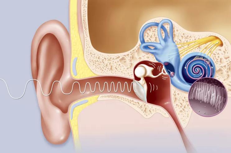
The Eustachian tube, also known as the auditory tube or pharyngeal tube, is a tube that connects the nasopharynx to the middle ear. This is part of the middle ear. In adults, the Eustachian tube has a length of about 35 mm (1.4 inches) and a diameter of 3 mm (0.12 inches). It is named after the sixteenth-century Italian anatomist Bartolomeo Eustaki.
In humans and other land animals, the middle ear (for example, the ear canal) is usually filled with air. However, unlike the open auditory canal, the air of the middle ear is not in direct contact with the atmosphere outside the body. The middle ear auditory tube provides a connection from the middle ear chamber to the back of the nasopharynx.
Usually the Eustachian tube is closed, but it opens when swallowed, and at positive pressure. In the case of take-off in an airplane, the ambient air pressure rises from higher (on the ground) to lower (in the sky). The air in the middle ear expands as the plane gains height and pushes into the back of the nose and mouth. On the way down, the volume of air in the middle ear decreases. So, a small vacuum is created. The active opening of the Eustachian tube is required to equalize the pressure between the middle ear and the surrounding atmosphere when the plane descends. The diver also experiences this pressure change, but at a faster rate. Active opening of the middle ear canal provides pressure equalization.
Composition
The Eustachian tube extends from the anterior wall of the middle ear to the lateral wall of the nasopharynx. This happens approximately at the level of the inferior nasal concha. It consists of a bone part and a cartilaginous part.
Bone part
The bone part (1/3) closest to the middle ear is made of bone and has a length of about 12 mm. It begins in the anterior wall of the tympanic cavity, below the septum of canalis musculotubarii, and, gradually tapering, ends at the angle of connection of the squamous and temporal parts of the temporal bone. The limb represents the serrated edge, which serves to attach the cartilage.
Cartilage
The cartilaginous part of the Eustachian tube is about 24 mm in length and is formed by a triangular plate of elastic fibrocartigal, the apex of which is attached to the edge of the medial end of the bone part of the tube. Its base lies directly beneath the mucous membrane of the nasal part of the pharynx, where the auditory tube of the middle ear provides the formation of an eminence - torus tubarius or pillows - behind the pharyngeal opening of the auditory tube.
The upper edge of the cartilage is twisted on its own, bent laterally so as to have a hook view in cross section; thus, a groove or groove is formed, which is open from below and from the side. This part of the channel is completed by a fibrous membrane. Cartilage lies in the groove between the malignant part of the temporal bone and the large wing of the sphenoid bone; this groove ends opposite the middle of the medial pterygoid plate.
Cartilage and bone sections of the tube are not in the same plane, with the first slope down slightly more than the second. The diameter of the tube throughout the body is heterogeneous. It is most in the hole for the pharynx, least of all - at the junction of the bone and cartilage parts. It is again enlarged in the direction of the tympanum. The narrowest part of the tube is called the isthmus.
The mucous membrane of the tube in front is continuous with the nasal part of the pharynx, and at the back with the cavity of the tympanic cavity. The middle ear auditory tube provides ciliary pseudostratified columnar epithelium. It is thin in the bony part, while in the cartilaginous part contains many mucous glands and near the pharyngeal opening is a significant amount of adenoid tissue, which Gerlach called the tube tonsil.
Muscle
There are four muscles associated with the function of the Eustachian tube:
- Levator veli palatini (innervated by the vagus nerve).
- Salpingopharyngeus (innervated by the vagus nerve).
- Tensor tympanic membrane (innervated by the mandibular nerve CN V).
- Great palatini tensor (innervated by the mandibular nerve CN V).
The auditory tube of the middle ear ensures that when swallowing, the membrane opens by reducing the veli palatini tensor and veli palatini levator, the muscles of the soft palate.
Development
The Eustachian tube originates from the ventral part of the first pharyngeal pocket and the second endodermal pocket, which forms a tubular depression during embryogenesis. The distal part of the tubular groove leads to the formation of a tympanic cavity, and the proximal tubular structure becomes the Eustachian tube. Thus, the middle ear auditory tube provides vibrations and helps the conversion of sound waves.
Functions
Pressure equalization. Under normal circumstances, the Eustachian tube of a person is closed, but it can open to let in a small amount of air and prevent damage by equalizing the pressure between the middle ear and the atmosphere. The pressure drop causes temporary conductive hearing loss due to reduced movement of the eardrum and ear bones. Various methods of ear cleaning, such as yawning, swallowing, or chewing gum, can be used to deliberately open the tube and balance the pressure. When this happens, people hear a quiet crack. This event is familiar to aircraft passengers, scuba divers or drivers in mountainous regions.

Devices that help in balancing the pressure include a special spray applied to the nose, creating inflation with positive air pressure. Some people learn to voluntarily “click” on their ears, together or separately, performing a pressure equalization procedure, opening their Eustachian tubes when pressure changes, for example, when lifting / lowering in an airplane, driving in the mountains, lifting / lowering an elevator, etc. d.
Some may even knowingly keep their Eustachian tubes open for a short period and even increase or decrease the air pressure in the middle ear. In fact, a “click” can be heard by raising the ear to another while making a click sound. This voluntary control can be first detected by yawning or swallowing, as well as by other means (see above). Those who develop such an ability may find that it can be done consciously, without power, even if there is no problem with pressure.
Drainage of mucus. Infections or allergies in the upper respiratory tract can cause the Eustachian tube or the membranes surrounding its opening to become swollen. They will retain fluid, which serves as a breeding ground for bacteria, causing ear infections, because the auditory tube of the middle ear allows mucus to drain from the middle ear. Ear infections are more common in children, since the tube is horizontal. It is shorter, which facilitates the penetration of bacteria, and also has a smaller diameter, which complicates the movement of fluid. In addition, the developing immune system in children and poor hygiene habits make them more prone to upper respiratory tract infections.