Everyone can perfectly imagine the human skeleton, thanks to the numerous photographs and drawings that each of us saw back in school. But do we know that the skeleton of an adult consists of a large number of different bones, each of which performs a specific function?
Human skeleton: what does it consist of?
The human skeleton is its support. It is not only capable of acting for the human body as a storehouse for its internal organs and systems, but it is also a place of attachment of its muscles.
With the help of the skeleton, a person is able to make various movements: walking, jumping, sitting, lying and much more. An interesting fact is that the human skeleton - the connection of bones - is formed in a child who is still in the womb. True, at first it was only
cartilaginous tissue, replaced in the process of his life with bone. In a baby, the bones have virtually no hollow space. It arises there in the process of human growth. One of the most important functions of the human skeleton is the formation of new blood cells that are produced by the bone marrow located in it. The peculiarity of the bones of the human skeleton is the preservation of a certain form throughout life (and therefore continuous growth and development). The list of bones of the human skeleton includes more than 200 items. Many of them are paired, while the rest do not form pairs (33-34 pieces). These are some of the bones of the sternum and skull, as well as the tailbone, sacrum, and vertebrae.
Human limb functions
It is very important to know that the process of evolution, i.e., the continuous development of man, left a direct imprint on the functioning of many of his bones.
The upper section of the human skeleton with its moving limbs is intended mainly for human survival in the world. With his hands, he is able to cook food, do homework, serve himself, etc. There are also bones of the lower extremities of a person. Their anatomy is so thought out that a person is able to stay upright. At the same time, they serve as the basis for movement and support for him. I want to note that the lower limbs are less mobile compared to the upper ones. They are more massive in weight and density. But along with this, their functions are very important for a person.
Human lower limb skeleton
Consider the human skeleton: the skeleton of the lower limb and upper limb is represented by a belt and a free part. In the upper section, these are the following bones: the chest girdle, shoulder blades and clavicle, the humerus and forearm bones, and the hand. The bones of the lower extremity of a person include: the pelvic girdle (or paired pelvic bones), thigh, lower leg, foot. The bones of a person’s free lower limb, as well as the belt, can support a person’s weight, which is why they are so important to him. After all, in fact, only with the help of these compounds can it be in an upright position.
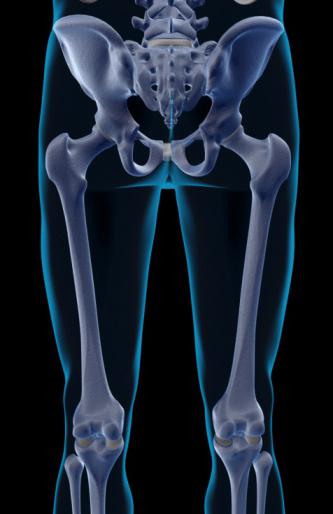
Pelvic girdle (paired pelvic bones)
The first component, which is the foundation that forms the bones of the lower limb, is the pelvic bone.
It is she who changes its structure after puberty of any adult. Until this age, they say that the pelvic girdle consists of three separate
bones (iliac, pubic and sciatic), interconnected by cartilage. Thus, they form a certain hollow where the femoral head is placed. The bone pelvis is formed by connecting the front of the same name bones. At the back, it is articulated with the help of the sacrum. As a result, the pelvic bones form a kind of ring, which is a repository for the internal organs of a person.
Femur and patella
The bones of the belt of the lower limb of a person are not as mobile as the rest of it, which is called the free lower limb. It consists of: thigh, lower leg and foot. The thigh, or femur, is a tubular bone. It is also the largest and longest of all the bones that endow the human body. In its upper part, the femur connects to the pelvic girdle with the help of the head and a long thin neck. Where the neck passes into the main part of the femur, there are two large tubercles on it. It is here that the bulk of the muscles of the lower extremities of the person are attached. Down the femur becomes thicker. There are also two elevations, thanks to which the hip connects, as a result, with the patella and lower leg. The patella is a rounded flat bone with the help of which the bend of the leg in the knee occurs. The bones of the lower limb of a person, namely the thigh and patella, carry the following functions: the place of attachment of the bulk of the muscles located on the legs, and the possibility of bending the legs.
Shin
The human tibia consists of two bones: the tibia and fibula. They are located next to each other.
The first one is quite massive and thick. From above, it connects with the outgrowths
(condyles) of the femur and the head of the fibula. Down the tibia turns on the one hand into the medial ankle, and on the other - is located directly under the skin. The fibula is smaller in size. But along the edges it is also thickened. Due to this, it connects from above to the tibia, and forms a lateral ankle from below. It is important that both components of the lower leg, which also represent the bones of the lower limb of a person, are tubular bones.
Human foot bones
The bones of the human foot are divided into three main parts: the bones of the tarsus, metatarsals and phalanges of the fingers. It is important to note that the foot is the free bones of the lower limb of a person. The first of them include seven bones, the main of which is the bone, called the talus and forming the ankle joint, and the calcaneus. Farther are the metatarsal bones. There are only five of them, the first of them is much thicker and shorter than the others. Toes are made up of bones called phalanges. The peculiarity of their structure is that the big toe contains 2 phalanges, the remaining toes - three pieces each.
Anatomy of the joints of the lower extremities of a person. Sacroiliac joint, pubic symphysis
I immediately want to say that all the joints of the lower limb are very large, compared with the joints of the upper limbs.
They have a large number of different ligaments, due to which the variety of movements that can be done with the help of a person’s legs is carried out. Bones and joints of bones of the lower limb were originally created in order to serve as a support to the human body and move it. Therefore, of course, they are reliable, strong and able to withstand heavy loads. Let's start with the highest joints. With their help, the pelvic bones are connected, and a human pelvis is formed. In front of such a joint is called pubic symphysis, and in the back - sacroiliac. The first is based on
pubic bones located towards each other
. Strengthening the pubic symphysis is formed due to a large number of ligaments. The sacroiliac joint is very strong and almost motionless. It is tightly bonded not only to the pelvic bones, but also to the lower spine using tight ligaments.
Human pelvis: large and small. Hip joint
It has already been described above that the bones of the belt of the lower extremity of a person are represented primarily by the pelvic bones. They, connecting with the help of the sacrum and pubic symphysis, form the pelvis. This, figuratively speaking, is a ring that protects all organs, vessels and nerve endings inside from external influences. Distinguish a large and small basin. In women, it is much wider and lower than in men. For the fair sex, everything is thought out to facilitate the birth process, so the pelvis has a more rounded shape and greater capacity.
Joints of bones of the lower limb are also represented by one of the most famous representatives of this group - the hip joint. What is he so famous for? Dislocation of the hip joint is the most famous defect in the development of the lower extremities, which can be detected literally a month after the birth of the baby. It is very important to do this on time, since this untreated diagnosis can bring a lot of trouble in adulthood. The hip joint consists of a basin of the pelvic bone and a femoral head. The joint under investigation has many ligaments, thanks to which it is strong and quite mobile. Typically, experienced orthopedists can diagnose an abnormality in the development of the hip joint in childhood by routine examination of the patient. Leaving the legs to the side in a supine position of 180 degrees is only possible with healthy hip joints.
Knee-joint
Imagine a human skeleton. Jointing of bones in the form of joints is necessary for a person to have strong bonding of bones and create maximum mobility of all his limbs. A great example of such a connection is the knee joint. Incidentally, it is considered the largest joint in the human body. And its structure is very complex: the knee joint is formed using the condyles of the femur, patella, and tibia. The entire joint is shrouded in reliable ligaments, which, along with ensuring the movement of the leg, hold it in position. Thanks to him, not only standing, but also walking is carried out. The knee joint can produce various movements: circular, flexion and extensor.
Ankle joint
This joint serves to directly connect the foot and lower leg. Around are numerous ligaments that provide a variety of movements and the necessary stability to the human body.
Metatarsophalangeal joints
The studied joints are interesting in their shape compared to other joints of the lower limb of a person. They look like a ball. Strengthening for them are the ligaments on the sides and on the sole of the foot. They can move, although their movements do not differ in variety: small leads to the sides, flexion and extension. Human foot consists of numerous (inactive) joints and ligaments. With their help, movement is carried out, while the human body has the necessary support. So, we can conclude that the bones of the belt of the lower extremity of a person are less mobile than the free bones of a similar section. But the functions of this are no less among those and others.
How do human limbs develop with age?
We all know that during the course of life certain skeletons of a person undergo certain transformations. The skeleton of the lower limb undergoes strong changes with age. Bones that develop on the basis of connective tissue have three stages of their change: connective tissue, cartilage and bone tissue.
Pelvic bone: it is laid during intrauterine development of the fetus. Formed cartilaginous layers between the pelvic bones are usually preserved until puberty. Then they become stiff.
Patella: ossification points may appear in a child by the age of 2 years, this completely happens somewhere in 7 years. Interestingly, the lower limbs in newborns grow much faster than in adults. The peak of such rapid growth falls on puberty: in girls - 13-14 years; boys - 12-13 years old.
Remember that the human skeleton is prone to various injuries in the form of injuries and even fractures. Since he is entrusted with the performance of such a large number of important body functions, he must be protected. Eat properly (food with sufficient calcium helps to strengthen the skeleton), lead an active lifestyle (physical education and sports), monitor your health (check for any abnormalities in the functioning of the skeleton by a competent specialist) - all this must be done by each person. And then you will meet your advanced age vigorous, healthy and cheerful.