To perform the functions of support, movement and protection in our body there is a system that includes bones, muscles, tendons and ligaments. All its parts grow and develop in close interaction. Their structure and properties are studied by the science of anatomy. The humerus is part of the free upper limb and, along with the bones of the forearm and the belt of the upper extremities - the scapula and collarbone - provides complex mechanical movements of the human hand. In this work, using the example of the humerus, we will study in detail the principles of the musculoskeletal system and find out how its structure is associated with the functions performed.
Features of tubular bones
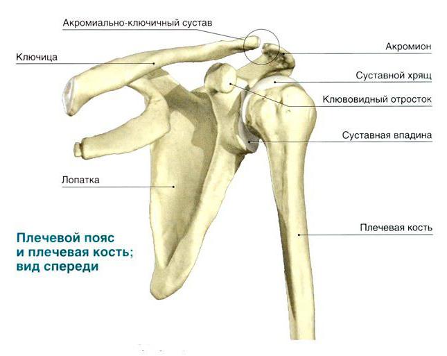
A trihedral or cylindrical shape is characteristic of the components of the skeleton - tubular bones, which distinguish elements such as the pineal glands (the edges of the bone) and its body (diaphysis). Three layers - the periosteum, the bone itself and the endoost - are part of the diaphysis of the humerus. The anatomy of the free upper limb is currently well understood. Epiphyses are known to contain spongy matter, while the central section is represented by bone plates. They form a compact substance. Long tubular bones have this appearance : the humerus, ulnar, and femur. The anatomy of the humerus, the photo of which is presented below, indicates that its shape in the best way corresponds to the formation of mobile joints with the bones of the belt of the upper extremities and forearm.
How do tubular bones develop?
In the process of embryonic development, the humerus, together with the entire skeleton, is formed from the middle embryonic leaf - the mesoderm. At the beginning of the fifth week of pregnancy, the fetus has mesenchymal areas called bookmarks. They grow in length and acquire the shape of the humerus tubular bones, the ossification of which continues after the birth of the baby. The humerus is covered by the periosteum from above. This is a thin shell consisting of connective tissue and having a branched network of blood vessels and nerve endings that enter the bone itself and provide its nutrition and innervation. It is located along the entire length of the tubular bone and forms the first layer of the diaphysis. As science has established anatomy, the humerus, covered by the periosteum, contains fibers of an elastic protein - collagen, as well as special cells called osteoblasts and osteoclasts. They are grouped near the central channel of Havers. With age, it becomes filled with yellow marrow.
Self-healing, repair and growth in the thickness of the tubular bones in the human skeleton is due to the periosteum. The anatomy of the humerus in the middle part of the diaphysis is specific. Here there is a tuberous surface to which the superficial deltoid muscle joins. Together with the belt of the upper extremities and the bones of the shoulder and forearm, it provides the lifting and abduction of the elbows and arms up, back and in front of you.
The value of the epiphyses of the tubular bones
The end parts of the tubular bone of the shoulder are called epiphyses, contain red bone marrow and consist of a spongy substance. Its cells produce blood cells - platelets and red blood cells. The epiphyses are covered with the periosteum, have bone plates and cords called trabeculae. They are located at an angle to each other and make up the inner skeleton in the form of a system of cavities, which are filled with hematopoietic tissue. As determined by the anatomy, the structure of the humerus at the junction with the scapula and the bones of the forearm is quite complex. The articular surfaces of the humerus have proximal and distal ends. The head of the bone has a convex surface, covered with hyaline cartilage and entering the cavity of the scapula. A special cartilaginous formation of the scapular cavity - the articular lip - serves as a shock absorber, mitigating tremors and shock when moving the shoulder. The capsule of the shoulder joint is attached at one end to the scapula, and at the other end to the head of the humerus, descending to its neck. It stabilizes the connection of the shoulder girdle and the free upper limb.
Features of the shoulder and elbow joints
As the human anatomy established, the humerus is a part of not only the spherical shoulder joint, but also one more - the complex ulnar. It should be noted that the shoulder joint is the most mobile in the human body. This is understandable, since the hand is the main instrument of labor operations, and its mobility is associated with adaptation to upright posture and exemption from participation in movement.
The elbow joint consists of three separate joints connected by a common joint capsule. The distal humerus connects to the ulnar, forming a blocky joint. At the same time, the head of the condyle of the humerus enters the fossa of the proximal end of the radial bone, forming a brachioradial movable joint.
Additional shoulder structures
The normal anatomy of the humerus includes the large and small apophyses, the tubercles from which the ridges extend. They serve as a place for attachment of the muscles of the shoulder. There is also a groove serving as a container for the biceps tendon. On the border with the body of the bone, the diaphysis, below the apophyses, is the surgical neck. She is most vulnerable to traumatic shoulder injuries - dislocations and fractures. In the middle of the bone body there is a tuberous area to which the deltoid muscle is attached, and behind it there is a furrow of a spiral shape into which the radial nerve is immersed. At the border of the epiphyses and the diaphysis, there is a section whose rapidly dividing cells determine the length of the humerus.
Disorders of the humerus
The most common injury is a fracture of the shoulder during a fall or a strong mechanical shock. The reason is that the joint does not have real ligaments and is stabilized only by the muscle corset of the upper extremity belt and an auxiliary ligament, which looks like a bundle of collagen fibrils. Damage to the soft tissues of the shoulder joint, for example, tendonitis and capsulitis, is quite common. In the first case, the tendons of the supraspinatus, infraspinatus, and small round muscles are damaged. Another disease occurs as a result of inflammatory processes in the joint capsule of the shoulder.
Pathologies are accompanied by tunneling pains in the arm and shoulder, limitation of the mobility of the shoulder joint when lifting the arms up, putting them behind the back, and abduction to the sides. All of these symptoms dramatically decrease a person’s performance and physical activity.
In this article, we studied the anatomical structure of the humerus and its relationship with the functions performed.