The heart is the most important organ in the human body. It is often compared with a motor, which is not surprising, because the main function of the heart is to constantly pump blood in the vessels of our body. The heart works 24 hours a day! But it so happens that it does not cope with its functions due to illness. Of course, it is necessary to monitor general health, including heart health, but in our time this is not obtained by everyone and not always.
A bit of ECG history
Even in the mid-19th century, doctors began to think about how to track work, identify deviations in time and prevent the terrible consequences of the functioning of a sick heart. Already at that time, doctors revealed that electrical phenomena were occurring in the contracting heart muscle , and began to conduct the first observations and studies on animals. Scientists from Europe began to work on creating a special apparatus or a unique technique for monitoring the work of the heart, and finally the world's first electrocardiograph was created. All this time, science did not stand still, thus, and in the modern world they use this unique and already improved apparatus, on which the so-called electrocardiography is produced, it is also called an abbreviated ECG. This technique of registration of biocurrents of the heart will be discussed in the article.
ECG procedure
Today it is an absolutely painless and accessible procedure for everyone. ECG can be done in almost any medical institution. Consult with your family doctor and he will tell you in detail what this procedure is for, how to take an ECG and where it can be taken in your city.
Short description
Consider the steps of how to take an ECG. The algorithm of actions is as follows:
- Preparing the patient for future manipulation. Laying it on the couch, the health worker asks to relax and not strain. Remove all unnecessary items, if any, and may interfere with the recording of the cardiograph. Exempt the necessary skin from clothing.
- Proceed with the application of electrodes strictly in a certain sequence and order of application of electrodes.
- Connect the device to work subject to all the rules.
- After the device is connected and ready for work, start recording.
- Remove the paper with the recorded electrocardiogram of the heart.
- The result of the ECG is given to the patient or doctor in his arms for subsequent decryption.
ECG preparation
Before you learn how to take an ECG, consider what actions you need to take to prepare the patient.
There is an ECG device in every medical institution, it is in a separate room with a couch for the convenience of the patient and medical staff. The room should be bright and comfortable, with an air temperature of +22 ... + 24 degrees Celsius. Since it is possible to correctly remove the ECG only if the patient is completely calm, such an environment is very important for this manipulation.
Place the subject on a medical couch. When lying down, the body easily relaxes, which is important for future recording of a cardiograph and for evaluating the work of the heart itself. Before applying electrodes for ECG, a cotton swab moistened with medical alcohol should be used to treat the required areas of the patient's arms and legs. Reprocessing of these places is carried out with physiological saline or a special medical gel intended for these purposes. The patient needs to remain calm during the recording of the cardiograph, to breathe smoothly, moderately, not to worry.
How to remove the ECG: electrode application
You need to know in what sequence you need to apply electrodes. For the convenience of the personnel conducting this manipulation, the inventors of the ECG apparatus identified 4 colors for the electrodes: red, yellow, green and black. They are superimposed precisely in this order and in no other way, otherwise an ECG will not be advisable. Confusing them is simply unacceptable. Therefore, the medical staff who works with the ECG apparatus undergoes special training with the subsequent passing of an exam and obtaining an admission or certificate allowing him to work with this particular apparatus. The health worker in the ECG office, according to his work instruction, must clearly know the places where the electrodes were applied and correctly perform the sequence.
So, the electrodes for arms and legs look like large clamps, but do not worry, the clamp is located on the limb completely painlessly, these clamps are of different colors and are applied to certain places of the body as follows:
- Red is the wrist of the right hand.
- Yellow is the wrist of the left hand.
- Green is the left leg.
- Black is the right leg.
Imposition of pectoral electrodes
Breast electrodes in our time are of different types, it all depends on the company of the manufacturer of the ECG apparatus itself . They are disposable and reusable. Disposable are more convenient to use, do not leave unpleasant traces of irritation on the skin after removal. But if there are no disposable ones, then reusable ones are used, they are similar in shape to hemispheres and have the property of sticking. This property is necessary for a clear statement in the right place with subsequent fixation at the right time.
A medical professional who already knows how to remove the ECG is located on the couch to the right of the patient in order to correctly place the electrodes. It is necessary, as already mentioned, to pre-treat the skin of the patient’s chest with alcohol, then with physiological saline or medical gel. Each chest electrode is marked. To make it clearer how to remove the ECG, the electrode application scheme is presented below.
We proceed to the application of electrodes on the chest:
- Previously, we find the 4th rib in the patient and put the first electrode under the rib, on which stands the number 1. In order for the electrode to successfully fall into place, you need to use its suction property.
- We also place the 2nd electrode under the 4th rib, only on the left side.
- Then we proceed to the imposition of not the 3rd, but immediately the 4th electrode. It is superimposed under the 5th rib.
- The electrode number 3 must be placed between the 2nd and 4th rib.
- The 5th electrode is mounted on the 5th rib.
- We put the 6th electrode at the level of the 5th, but a couple of centimeters closer to the couch.
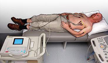
Before turning on the apparatus for recording ECG, we once again check the correctness and reliability of the applied electrodes. Only then can the electrocardiograph be turned on. Before this, you need to set the speed of the paper and configure other indicators. During recording, the patient should be in a state of complete rest! At the end of the machine, you can remove the paper with a cardiograph and release the patient.
We remove the ECG for children
Since there are no age restrictions for conducting an ECG, children can also take an ECG. This procedure is done in the same way as adults, starting from any age, including the neonatal period (as a rule, at such an early age, ECGs are done solely to eliminate suspicions of heart disease).
The only difference between how to remove the ECG for an adult and a child is that a special approach is needed for the child, he needs to explain everything and show, calm down if necessary. The electrodes on the body of the child are fixed in the same places as in adults, and must correspond to the age of the child. How to apply electrodes for ECG to the body, you are already familiar. In order not to excite the little patient, it is important to ensure that the child does not move during the procedure, support him in every possible way and explain everything that happens.
Very often, pediatricians, when prescribing an ECG, recommend additional tests for children , with physical activity or with the appointment of a particular drug. These tests are carried out in order to identify abnormalities in the work of the child’s heart in time, correctly diagnose a particular heart disease, prescribe treatment in time, or dispel the fears of parents and doctors.
How to remove an ECG. Scheme
In order to read correctly the recording on a paper tape that the ECG apparatus gives us at the end of the procedure, it is certainly necessary to have a medical education. The record should be carefully examined by a physician - therapist or cardiologist in order to timely and accurately establish a diagnosis for the patient. So, what can we tell an incomprehensible curve line consisting of teeth, individual segments at intervals? Let's try to figure it out.
The recording will analyze how regular the heart contractions are, reveal the heart rate, the focal point, the conductive ability of the heart muscle, the definition of the heart in relation to the axes, the state of the so-called cardiac teeth in medicine.
Immediately after reading the cardiogram, an experienced doctor will be able to diagnose and prescribe treatment, or give the necessary recommendations, which will significantly speed up the healing process or save from serious complications, and most importantly, an ECG performed on time can save a person’s life.
It must be taken into account that the cardiogram of an adult is different from the cardiogram of a child or a pregnant woman.
Do pregnant women have an ECG?
In what cases is a pregnant woman prescribed to undergo an electrocardiogram of the heart? If at the next appointment with the obstetrician-gynecologist the patient complains of pain behind the sternum, shortness of breath, large fluctuations in controlling blood pressure, headaches, fainting, dizziness, then most likely an experienced doctor will prescribe this procedure in order to reject bad suspicions in time and avoid unpleasant health consequences of the future mother and her baby. There are no contraindications for ECG during pregnancy.
Some recommendations before the planned ECG procedure
Before taking the ECG, the patient must be instructed on what conditions must be met the day before and on the day of removal.
- The day before, it is recommended to avoid nerve stress, and the duration of sleep should be at least 8 hours.
- On the day of delivery, you need a small breakfast of food that is easily digested, a prerequisite is not to overeat.
- Exclude products that affect the functioning of the heart for 1 day, for example, strong coffee or tea, spicy seasonings, alcoholic beverages, and smoking.
- Do not apply cream and lotions on the skin of hands, feet, chest, fatty acids which can subsequently worsen the conductivity of the medical gel on the skin before applying electrodes.
- Absolute calm is necessary before passing the ECG during the procedure itself.
- Be sure to exclude physical activity on the day of the procedure.
- Before the procedure itself, you need to sit quietly for about 15-20 minutes, breathing is calm, uniform.
If the subject has severe shortness of breath, then he needs to undergo an ECG not lying down, but sitting, since it is in this position of the body that the apparatus can clearly record cardiac arrhythmia.
Cardiologists recommend that all people undergo the procedure, without exception, after 40 years once a year.
Of course, there are conditions in which it is absolutely impossible to conduct an ECG, namely:
- In acute myocardial infarction.
- Unstable angina pectoris.
- Heart failure.
- Some types of arrhythmias of unclear etiology.
- Severe forms of aortic stenosis.
- Pulmonary embolism syndrome (pulmonary embolism).
- Stratification of the aortic aneurysm.
- Acute inflammatory diseases of the heart muscle and pericardial muscle.
- Severe infectious diseases.
- Severe mental illness.
ECG with a mirror arrangement of internal organs
Mirror arrangement of internal organs implies their arrangement in a different order, when the heart is not on the left, but on the right. The same applies to other organs. This is a fairly rare occurrence, but it does occur. When a patient with a specular arrangement of internal organs is prescribed to undergo an ECG, he must warn the nurse who will perform this procedure about his features. Young specialists working with people with a mirror arrangement of internal organs, in this case, the question arises: how to remove the ECG? On the right (the removal algorithm is basically the same), the electrodes are located on the body in the same order that ordinary patients would put on the left.
Take care of your health and the health of your loved ones!