The human esophagus is a muscle narrow tube. It is the channel through which food moves. The length of the human esophagus is about 25 centimeters. Next, we consider this section in more detail. We will find out where the person’s esophagus is, what tasks it implements. The article will also talk about the components of this department, as well as about some of the most common pathologies of the organ.
general information
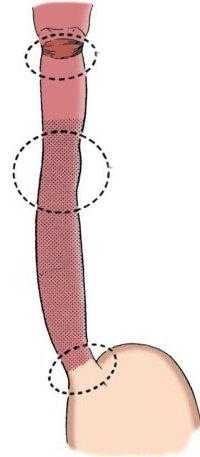
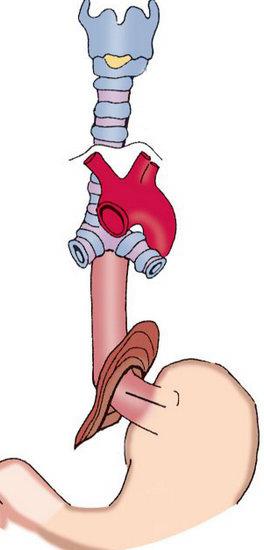
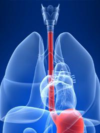
The esophagus and human stomach are two successively located sections of the gastrointestinal tract. The second is below. The first is located in the zone from the 6th cervical to 11 thoracic vertebrae. What is the structure of the human esophagus? It consists of three parts. The department includes the abdominal, thoracic and cervical zones. For clarity, the scheme of the human esophagus will be presented below. In the department there are also sphincters - upper and lower. They play the role of valves that provide unidirectional passage of food through the digestive tract. Sphincters prevent the penetration of aggressive contents from the stomach into the esophagus, and then the pharynx and oral cavity. There are also constrictions in the department. There are five of them. Two constrictions - pharyngeal and diaphragmatic - are considered anatomical. Three of them - bronchial, cardiac and aortic - are physiological. Such, in general, is the structure of the human esophagus. Next, we consider in more detail what the shells of an organ are.
Human esophagus anatomy
The department has a wall built of mucous, submucosal, as well as adventitial and muscle layers. The latter in the upper part of the department is formed by striated fibers. In approximately 2/3 (counting from above), the structures are replaced by smooth muscle tissue. In the muscular membrane there are two layers: the inner circular and the longitudinal outer. The mucous membrane is covered by a squamous stratified epithelium. In the thickness of this shell there are glands that open into the lumen of the organ. The mucous has a skin type. Flat layered epithelium lies on fine-fibrous connective fibers. This intrinsic layer of the membrane consists of collagen structures. Connective tissue cells and reticulin fibers are also present in the epithelium. The intrinsic layer of the membrane enters into it in the form of papillae. In general, the anatomy of the human esophagus is quite simple. However, it is important not so much as the tasks that are implemented in this department of the gastrointestinal tract.
Human Esophagus Functions
This department performs several tasks. The functions of the human esophagus include ensuring the promotion of food. This task is achieved through peristalsis, muscle contraction, changes in pressure and gravity. Also, mucus is secreted in the walls of the department. It is saturated with a food lump, which facilitates its penetration into the cavity of the stomach. Also, the channel’s objectives include providing protection against the return of contents to the upper gastrointestinal tract. This function is realized thanks to the sphincter.
Business disruption
Comparing the prevalence of pathologies of the esophagus and stomach, one can notice the following: the former are detected much less frequently. Normally, the food taken passes without delay. It is believed that the human esophagus is less susceptible to certain irritations. In general, this department is quite simple in its structure. However, there are some nuances in its structure. Today, experts have studied most of the existing congenital and acquired malformations of the department. More often than not, doctors diagnose abnormal anatomy of the sphincter connecting the stomach to the esophagus. Another fairly common defect is difficulty swallowing. In this pathological condition, the diameter of the human esophagus is reduced (normally it is 2-3 cm).
Symptoms of Disease
Often the pathology of the esophagus is not accompanied by any manifestations. Nevertheless, violations in his work can lead to quite serious consequences. In this regard, it is necessary to pay attention even to seemingly minor symptoms. If any prerequisites are found, then you should immediately visit a doctor. Among the most common symptoms of esophageal pathologies should be noted:
- Heartburn.
- Burping.
- Pain in the epigastrium.
- Difficulty eating.
- Lump in throat.
- Soreness in the esophagus during eating.
- Hiccups.
- Vomiting
Spasm
In some cases, difficulty in passing food is associated with spastic contractions of the muscles of the esophagus. Usually this condition is noted in young people. More prone to the development of spasm of the face, subject to excitability and characterized by instability of the central nervous system. Often the condition occurs in conditions of stress, rapid absorption of food, general nervousness. At a high rate of consumption of products, the human esophagus is mechanically irritated. As a result, a spasm develops at a reflex level. Often muscle contraction is noted at the junction of the esophagus and stomach. In this case, cardiospasm occurs. Consider this condition in more detail.
Cardiospasm
This condition accompanies the expansion of the esophagus. This anomaly is characterized by a giant increase in its cavity with morphological changes in the walls against the background of a sharp narrowing of its cardial part - cardiospasm. Expansion of the esophagus can develop due to a variety of external and internal pathogenic factors, impaired embryogenesis, neurogenic dysfunctions leading to atony.
Causes of cardiospasm
The pathological condition is supported by traumatic injury, an ulcer, a tumor. A provoking factor for further development is the effect of toxic compounds. These, first of all, include couples in hazardous industries, alcohol, tobacco. Increases the likelihood of developing cardiospasm of esophageal stenosis due to a lesion in the presence of typhoid, scarlet fever, syphilis and tuberculosis. Among the provoking factors, a special place is occupied by various pathologies of the diaphragm. These, in particular, include sclerosis of the hole. Subphrenic phenomena in the organs of the abdominal cavity also have a negative effect. In this case, we are talking about aerophagia, gastritis, gastroptosis, peritonitis, splenomegaly, hepatomegaly. The provoking factors include supradiaphragmatic processes. Among them, in particular, aortic aneurysm, aortitis, pleurisy, and mediastinitis are distinguished. Neurogenic factors include damage to the nerve peripheral apparatus of the esophagus. They can be caused by some infectious pathologies. For example, measles, typhus, diphtheria, scarlet fever, meningoencephalitis, influenza, and poliomyelitis can become the cause. Also, provoking factors include poisoning with toxic compounds in the workplace and at home (lead, alcohol, arsenic, nicotine). Changes in the esophagus leading to gigantism are likely to develop at the stage of embryonic laying. Subsequently, this is manifested by sclerosis, thinning of the walls.
Achalasia
This disorder is neurogenic. With achalasia, a violation of the functions of the esophagus is noted. With pathology, disorders in peristalsis are observed. The lower sphincter, which acts as a locking mechanism between the esophagus and stomach, loses its ability to relax. At present, the etiology of the disease is unknown, but experts speak of a psychogenic, infectious and genetic predisposition. Usually pathology is detected at the age of 20 to 40 years.
Burns
They arise if certain chemical compounds penetrate the human esophagus. According to statistics, of the total number of people who received burns in this section of the gastrointestinal tract, approximately 70% are children under ten years of age. Such a high percentage is due to the oversight of adults and the curiosity of the kids, provoking them to taste many things. Often, adults get a burn of the esophagus when penetrating caustic soda, concentrated acid solutions. Less common are cases of exposure to lysol, phenol. The degree of damage is determined in accordance with the volume and concentration of the compound that has got inside. At 1 tbsp. damage to the surface layer of the mucosa is noted. The second degree is characterized by lesions in the muscles. Esophagus burn 3 tbsp. accompanied by damage in all layers of the department. In this case, not only local symptoms appear, but also general signs: intoxication and shock. After a burn, 2-3 tbsp. cicatricial changes in the tissues are formed. The main symptom is a burning sensation in the mouth, throat and behind the sternum. Often, a person who has taken a caustic solution immediately experiences vomiting, and swelling of the lips may appear.
Foreign body
Sometimes objects not intended for digestion enter the human esophagus. As foreign bodies, unexposed chunks of food can act. As practice shows, the presence of foreign elements is diagnosed quite often. A foreign body may appear in the esophagus due to eating foods too fast, laughing or talking while eating. Often bones of fish or chicken are found in this section. The appearance of a foreign object is characteristic of people who have the habit of holding something inedible in their mouths constantly (paper clips, cloves, matches, etc.). As a rule, objects with a pointed end are introduced into the wall of the organ. This can trigger an inflammatory process.
Ulcer
Such a pathology can be caused by insufficient cardia, which provokes the penetration of gastric juice into the esophagus. He, in turn, has a proteolytic effect. Often an ulcer is accompanied by damage to the stomach and duodenum or hernia in the esophagus of the diaphragm. Typically, single lesions are found on the walls, but in a number of cases multiple expressions are diagnosed. Several factors contribute to the development of an esophageal ulcer. Pathology may result from surgery, hernia, or peristalsis. The main symptoms are constant heartburn, pain behind the sternum, belching. When taking food and after it, these manifestations become more intense. Periodically arising regurgitation of acidic contents from the stomach also acts as a characteristic sign.
Atresia
This defect is considered quite severe. For pathology, the blind end of the upper esophagus is characteristic. At the same time, its lower segment communicates with the trachea. Often, against the background of atresia of the esophagus , other malformations in the development of various systems of the body are revealed. Causes of pathology are considered abnormalities in the intrauterine formation of the fetus. If at 4 or 5 weeks of development the embryo will be affected by harmful factors, then later the wrong formation of the esophagus may begin.