Magnetic resonance imaging (MRI) of the lumbosacral region of the spine is an innovative method of radiation diagnostics, which is based on the characteristics of the atoms of the human body to respond to magnetic effects. In it, to obtain images of the lumbar (lower) part, a high power electromagnetic field and radio frequency pulses are used. Special sensors of the tomograph allow not to shift a person in the case when it is also necessary to remove the upper zone of the spine. X-ray radiation is not used during the examination, therefore the procedure is not dangerous for the patient’s health and can be repeated many times if necessary.
MRI of the lumbosacral spine is performed in the sagittal and transverse projections at the lesion levels. It has the ability to simultaneously see on the monitor multiple slices - images that are obtained with MRI. The optimum result is achieved with a section thickness of 3 to 4 mm (there should be no gaps between them). In one examination session, from several tens to hundreds of sections are produced. Then they are stored in the computer memory or printed on film. Today, MRI of the lumbar spine is considered the most accurate method of research and is called the "gold standard" in diagnosis.
When is MRI indicated at the lumbar level?
The study helps to identify pathological changes in the spine, as well as in the tissues adjacent to it. When using a magnetic resonance imager, a diagnosis is possible at an early stage of the disease.
This makes it possible to timely proceed with the treatment of pathologies associated with the spine and obtain a successful cure result. MRI of the lumbosacral spine is prescribed in such cases:
- violation of the sensitivity of the lower extremities, weakness in the legs;
- non-treatable pain in the lumbar region;
- trauma in the lower back and sacrum, for example, a fracture or instability of the spine;
- symptoms of a malignant tumor together with pain at the level of the lumbar spine;
- a history of cancer and metastases of malignant cells in the lower back;
- congenital malformations and abnormalities in the development of this spine;
- infections and abscesses of the spinal cord and bones;
- disturbances in the urination process.
What does MRI of the lumbosacral spine show?
During the examination, the doctor can identify abnormalities. MRI allows you to analyze the condition of the spinal cord in general, the intensity of the chemical and physical processes in it, and also to examine in detail the state of the vessels.
In cases where a sharper image of the abnormal part of the tissue is required, an MRI of the spine is performed using a contrast medium. It allows for clearer visualization, for example, helping to distinguish injuries and diseases from past surgical scars. But more often the procedure with contrast is used to study or detect tumors of various nature. MRI of the lumbosacral spine determines:
- problems that arise in the intervertebral discs, such as pinched nerves, ruptured discs, inflammation of the sciatic nerve;
- an area of the spine with an abnormal narrowing of the canals (stenosis), which is a possible indication for surgery;
- benign formations (hemangiomas) developing in the body of the vertebrae;
- the effect on the nerves and bones of the spine of malignant tumors of other nearby organs or their metastases;
- intervertebral hernia and disc protrusion in this area;
- arthritis (inflamed joints), as well as bone loss;
- stenosis of the spinal canal (its narrowing);
- spondylitis (degenerative changes in the vertebrae);
- areas with poor blood supply;
- infectious processes at the level of the lumbar;
- nerve damage due to injury or illness, for example, multiple sclerosis;
- congenital malformations of the spine.
Spinal cord examination
The spinal cord includes the nuclei of the peripheral and central nervous systems, so pathological processes in it can lead to serious diseases, up to disability and death. MRI of the spinal cord and spine of the lumbosacral allows neurologists and neurosurgeons to timely diagnose and cure a number of diseases that would become incurable without the capabilities of a magnetic resonance imager.
The image shows the image of a long light gray strand surrounded by white fluid - this is the spinal cord. It is located in the background of the vertebral bodies. The survey assesses its structure and size. For example, in the absence of pathologies, the spinal cord has clear and even edges, is located in the center of the spinal canal, and with an increased size, an oncological process is possible. Magnetic resonance diagnostics of the spinal cord is prescribed in such cases:
- detection of focal lesions, malignant neoplasms of the spinal cord, as well as its membranes;
- suspected injury, malignant lesions of the spine, as well as other pathologies of vertebral origin;
- determination of syringomyelia by the method of assessing its cerebrospinal fluid spaces;
- analysis of the condition of the spinal cord after surgical interventions in this area.
Indications for MRI in the coccyx
Coccyx MRI is a reliable non-radiation diagnostic method that allows you to assess the condition of the lower zone of the spine and the coccygeal region and determine various pathologies at the initial stage of occurrence.
This study is extremely rare in isolation. Usually, if anomalies in this area are suspected, they are sent to an MRI of the lumbosacral spine and tailbone, sometimes also together with the pelvic organs. The optimal area for examination is prescribed by a doctor, depending on the symptoms. Indications for coccyx MRI:
- any traumatic injuries in the sacrococcygeal region;
- congenital or acquired abnormalities in the development of the sacrum and tailbone, namely lumbarization of the vertebrae of the sacrum, cysts and coccygeal passages;
- suspected presence of hemangiomas, cysts or malignant tumors, as well as metastasis to the tailbone or sacrum;
- pains of any nature, localized in the lower back;
- circulatory disorders and weakening of reflexes in the lower extremities, as well as their paralysis;
- suspected pathology of nerve roots ( ponytail syndrome );
- fuzzy or questionable results from other surveys in this area.
What diagnoses MRI in the examination of the thoracic region?
This study is most often used to assess the structure of the spine from the inside. Magnetic resonance imaging makes it possible to see the intervertebral discs, the spinal canal, muscles, other soft tissues, the upper region of the lumbar region and the lower zone near the cervical spine, as well as the vertebral bodies and their processes.
MRI of the thoracic spine provides the opportunity to diagnose:
- osteochondrosis;
- narrowing of the spinal canal, as well as openings between the spinal nerves (stenosis);
- malignant neoplasms and their metastases in the thoracic region;
- acute circulatory disorders;
- abnormalities in the development of this zone of the spine;
- intervertebral hernia and protrusion of the spine;
- any injuries in this area;
- demyelinating diseases ( acute disseminated encephalomyelitis or multiple sclerosis);
- the presence of vascular malformations in the spinal cord.
Conducting research and contraindications to it
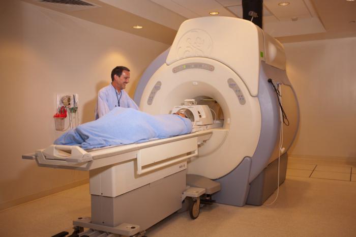
The examination does not require special preparations. Before it begins, it is necessary to remove all objects that contain metal. The procedure is carried out in a horizontal position. To get high-quality images, a person must be in a stationary state. If the patient is a child who cannot be without movement, then it is possible to use anesthesia, sleeping pills or sedatives to undergo MRI of the lumbosacral spine. Contraindications are almost identical for all types of magnetic resonance diagnostics:
- electronic devices, such as a pacemaker in a patient;
- the presence of foreign bodies with metal in the composition: implants, plates, pins, structures for osteosynthesis and others;
- inadequate mental behavior;
- claustrophobia;
- pregnancy up to 12 weeks, since the effect of a magnetic field on fetal development has not yet been adequately studied;
- when you enter a contrast agent, an allergic reaction to it is added;
- Nursing mothers are prohibited from applying the baby to the breast for 48 hours after the procedure with contrast.
Where is the best MRI scan in Lublin?
MRI of the lumbosacral spine will help to identify various pathological processes in any areas of the spine, vertebrae, spinal cord, soft tissues. In Lublin, there is a medical center that offers to undergo this study on a modern high-quality magnetic resonance imager.
Professional staff of the center and the latest generation of equipment guarantee high accuracy and maximum comfort of the examination procedure.
Price for MRI
The volume of work performed, the use of contrast medium, the area of the scan area, additional services, and tasks - all these factors affect the cost of MRI. Research prices for different areas may vary. On average, MRI of the lumbosacral region of the spine without the use of contrast medium is in the range of 4000-5000 rubles, and with contrast - about 9000 rubles. You can make an appointment with a specialist of a medical center for a more detailed consultation on this issue.
Finally
MRI of the lumbosacral spine is a painless and highly informative diagnostic method with a number of advantages over other methods. It is absolutely harmless, because the patient is not exposed to radiation exposure. MRI has no analogues for the diagnosis of not only protrusions of the intervertebral discs, but also their hernias. Magnetic resonance imaging makes it possible to examine the spine simultaneously with soft tissues without the use of a contrast medium, in contrast to devices with x-ray diagnostic methods. Carrying out MRI in the chest area is the most informative way to assess the condition of soft tissues, to identify the localization and size of tumors, as well as to study the cartilage surface of joints, tendons or muscles.