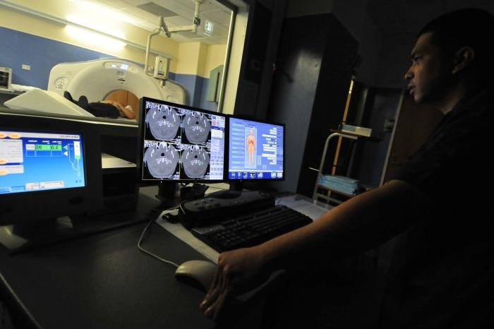
Computed tomography of the brain is a diagnostic study that allows you to take pictures of the inside of the head using x-rays and computer technology. During the procedure, the patient is placed on the table, and his head is placed in a tomograph, which is a large machine in the form of a box with a hole in the center. The scanner, as it were, transmits rays in a special way, giving out pictures of the skull (it can be tilted to take photos from different positions). Then all the images are saved on the computer, and if necessary can be printed.
Computed
tomography of the brain can give a more complete picture of brain tissues and structures than standard images, thereby providing additional information about injuries and diseases. It is carried out if necessary:
- determine the cause of confusion, headache and dizziness, paralysis, numbness, which may be evidence of a tumor or brain injury, bleeding in the head, rupture of the aneurysm;
- find damage caused by a stroke;
- check how successful the surgical removal or treatment of a brain tumor has been;
- identify structural brain abnormalities (such as hydrocephalus);
- detect blood clots in the brain.
Head tomography can also be prescribed for other purposes, for example, to plan radiation therapy for brain cancer. Or as a guide for a biopsy. Before the procedure, the doctor explains to the patient its essence and offers to ask questions of interest. Tell your doctor if you have conditions such as pregnancy, allergies to any medications, heart disease, diabetes, kidney problems, asthma!
Computed tomography of the brain is as follows. First, the patient must remove clothing and jewelry that interfere with the procedure. During the study, the head will be immobilized to prevent its movement. The diagnostics technologist will constantly monitor its progress. When computed tomography of the brain is performed, the patient may hear clicks, which is normal. Information obtained as a result of scanning is transferred to a computer, which transforms it into images. Then these images are interpreted by a radiologist.
Complete study results are usually prepared within a few days. But the radiologist can immediately after the procedure discuss with the patient some aspects that were revealed by computed tomography of the brain. The cost of such diagnostic testing, by the way, is quite acceptable. And it varies in the range from three to six thousand rubles. At the same time, the benefits of CT are undeniable. This painless and accurate method allows you to make very detailed images of bone and brain tissue, as well as to identify bleeding and internal damage as soon as possible. In many cases, this is priceless!