A head injury, the consequences of which can be completely different (up to a fatal outcome), is one of the common causes of disability in middle and young age. About half of all cases are TBI. According to statistics, about 25-30% of all injuries are brain damage. These cases account for more than half of the deaths. Further in the article a classification of injuries will be presented, some of them will be described.
General information
Traumatic brain injury refers to damage to the bones of the skull or soft tissues. The latter, for example, include the meninges, nerves, blood vessels, and others. Head injuries are divided into several groups. Let's consider some of them in more detail.
Injury classification
Damage may be exposed. In this case, the aponeurosis and the skin are injured. The bone or tissue lying more deeply acts as the bottom of the wound. Penetrating injury is characterized by damage to the hard shell of the brain. As a special case, otliquorrhea due to a fracture of bones at the base of the skull can be considered . A closed head injury may also occur . In this case, the skin can be damaged, and the aponeurosis maintains its integrity. The following groups are also distinguished:
- Tremors. These are head injuries that are not characterized by persistent disturbances in the brain. All manifestations of the condition after a while (usually a few days) disappear on their own. With more persistent symptoms, a more severe head injury occurs with possible brain damage. The main criteria for assessing the condition is the duration of the concussion (from seconds to several hours) and the subsequent depth of the state of amnesia and loss of consciousness. Among non-specific symptoms, vomiting, nausea, cardiac abnormalities, blanching of the skin should be noted.
- Compression of the brain by the focus of the bruise, air, foreign body, hematoma.
- Subarachnoid hemorrhage.
- Diffuse axonal lesion.
In practice, a lot of combined cases have been recorded. For example, hematoma compression and contusion, contusion with subarachnoid hemorrhage and compression, diffuse damage and contusion, etc. may be combined. Often, damage occurs due to a facial injury.
Brain injury
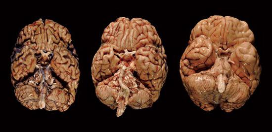
It occurs against a background of a head injury. A bruise is a violation of the integrity of the substance of the brain in a certain limited area. As a rule, such a region arises at the point of application of force. However, there are cases when a bruise appears on the opposite side (from a counter-shock). Against the background of this condition, part of the brain tissue, blood vessels, histological cellular connections are destroyed with the subsequent formation of traumatic edema. The area of such lesions is different. Of particular danger is such a head injury in a child.
Mild
Such head injuries are characterized by a loss of consciousness for a short period - up to several tens of minutes. After graduation, complaints of nausea are typical. Also, the patient hurts and is dizzy. Vomiting may occur, in some cases repeated. In some cases, moderate bradycardia is observed - a decrease in the frequency of heart contractions to 60 or less per minute. The patient may have con-, retro- and anterograde amnesia - a memory impairment in the form of a loss of the ability to preserve and reproduce previously acquired knowledge. After a mild head injury, tachycardia is noted (an increase in the heart rate to 90 beats / min). In some patients, pressure may rise. In this case, body temperature and respiration, as a rule, remain unchanged. As for neurological symptoms, the manifestations are usually mild. So, the patient may have weakness, drowsiness, clonic nystagmus (biphasic rhythmic involuntary eye movements ). There is also a slight anisocoria, meningeal symptoms, pyramidal insufficiency. These manifestations usually regress 2-3 weeks after a head injury.

Characterization of Violations
Against the background of a bruise, a coarse damage to the brain substance is microscopically detected. It manifests itself as sites of local swelling, cortical puncture bruising, probably in combination with subarachnoid limited hemorrhage. It, in turn, is due to rupture of the pial vessels. Blood with subarachnoid hemorrhage penetrates under the arachnoid membrane and spreads along the basal cisterns, cracks and furrows of the brain. It can be local or fill the entire space with the formation of clots. The condition is developing quite sharply. The patient suddenly feels a “blow to the head”, photophobia, vomiting, very severe headache quickly appear. Repeated generalized convulsions are likely. Usually the condition is not accompanied by paralysis. However, meningeal symptoms are likely. In particular, stiff neck muscles (tilting the head cannot touch the sternum of the patient’s chin) and Kerning’s symptom (the leg bent in it and the hip joint cannot be bent) can be noted. In the presence of meningeal symptoms, irritation of the meninges with poured blood takes place.
Moderate bruise
This head injury is characterized by a longer shutdown of consciousness (up to several hours). The patient has severe amnesia. The following signs of head trauma are also observed: severe headache, repeated vomiting, mental disorders. Transient disturbances in vital functions are likely. In particular, tachycardia or bradycardia, increased pressure, tachypnea (shallow rapid breathing without disturbing the rhythm and patency of the paths), subfebrile condition (body temperature rises to 37-37.9 degrees) can be noted. Stem and sheath symptoms, dissociation of tendon reflexes and muscle tone, bilateral pathological manifestations are common. Focal symptoms are quite clear. Her character is determined by the localization of the bruise. There are oculomotor and pupil disorders, speech disorders, sensitivity, paresis of limbs and others. These symptoms gradually smooth out over the course of three to five weeks, as a rule. However, in some cases, the described clinical picture persists for a sufficiently long time. With a moderate bruise, fractures in the bones of the base and fornix of the skull, extensive subarachnoid hemorrhage are often found. On CT, focal changes are detected in the form of small high-density inclusions or a homogeneous moderate increase in density. This corresponds to minor hemorrhages in the area of the bruise or hemorrhagic impregnation of brain tissue without gross destruction.
Severe head injury
In this case, intracerebral hematomas are noted in both frontal lobes in the form of limited blood clusters with various injuries with rupture of blood vessels. In this case, a cavity is formed that contains coagulated or liquid blood. A bruise is severely characterized by a prolonged loss of consciousness (up to several weeks). Often marked motor agitation is noted. Disorders of vital functions in the body are also noted. However, in comparison with the average degree, in severe they are more pronounced. So, for example, there is a disorder of respiratory function with impaired patency of the paths and rhythm. The patient has hyperthermia, the dominance of primary-stem neurological symptoms. In particular, swallowing disorders, floating eye movements, ptosis or mydriasis, paresis of the eye, decerebral rigidity, nystagmus, increased or inhibited reflexes of the mucous membranes, skin, tendons and so on are detected. Neurological symptoms in the initial period (in the first hours or days) prevail over focal hemispheric manifestations. The patient may have paresis of limbs, subcortical disorders of muscle tone, and more. In some cases, focal or generalized epileptic seizures are likely . Regression of focal manifestations occurs rather slowly. What is the danger of such a head injury? The consequences can be quite serious. Often expressed residual effects are observed, mainly in the mental and motor sphere.
CT scores
In severe trauma, in the third part of cases focal lesions in the brain are observed in the form of heterogeneous areas of increased density. In this case, an alternation of zones is observed. High and low density areas are distinguished. In the most severe course of the condition, the destruction of the brain substance goes deeper and can reach the ventricular system and subcortical nuclei. Observations of the dynamics show a gradual decrease in the volume of compacted areas, their merging and transformation into a more homogeneous mass. This happens on the 8th or 10th day after the incident. The regression of the volumetric effect of the pathological substrate is slower, which indicates the presence of unresolved clots and crushed tissue in the focus of the injury. At this point, they become equally dense relative to the surrounding edematous medulla. Disappearance after 30-40 days. the volumetric effect indicates the resorption of the substrate and the formation of areas of atrophy or cystic cavities instead.
Damage to the structures of the posterior cranial fossa
This lesion is considered the most severe of all head injuries. The condition is characterized by the following symptoms: depression of consciousness and a combination of stem, cerebellar, meningeal and cerebral symptoms, due to rapid compression and disturbances of cerebrospinal fluid circulation.
Therapeutic measures for bruising
Regardless of the degree of damage, medical assistance should be provided to the patient. In case of head injury, the victim must be transported to the hospital as soon as possible. To make an accurate diagnosis, radiography and CT are shown. The patient needs bed rest. Its duration with a mild degree is 7-10 days., With an average of up to 14 days. In case of severe head injury, resuscitation measures must be taken. They begin in the prehospital period and continue under stationary conditions. To normalize breathing, it is necessary to ensure free patency in the upper respiratory tract - they release them from mucus, blood, and vomit. An air duct is inserted, a tracheostomy is performed (dissection of the tracheal tissue and insertion of a cannula or the formation of a permanent opening - stoma). Inhalation using an oxygen-air mixture is also used. If necessary, mechanical ventilation is used.
Concussion therapy
If it is established that the patient has a head injury, treatment should be carried out in a neurosurgical hospital. With a concussion, a five-day bed rest is indicated. In the absence of complications, the patient can be discharged on the 7-10th day. At the same time, he is prescribed outpatient treatment, the duration of which is up to 14 days. Drug therapy for concussion is aimed at stabilizing the functional state of the brain, eliminating pain, insomnia, anxiety. As a rule, the range of prescribed medications includes sleeping pills, sedatives and painkillers. As analgesics, drugs such as Baralgin, Pentalgin, Maksigan, Sedalgin and others are used. With dizziness, Cerucal may be prescribed. Medications such as Valocordin and Corvalol are considered sedatives. and others containing phenobarbital, infusions of herbs (motherwort, valerian) are used.
Tranquilizers are also recommended. For example, they include such funds as Rudotel, Nozepam, Phenazepam, Sibazon, Elenium and others. In addition to symptomatic therapy, metabolic and vascular treatment is prescribed. It contributes to a faster and more complete restoration of impaired brain functions, and prevents various post-commotion symptoms. The appointment of cerebrotropic and vasotropic therapy is allowed 5-7 days after injury. It is advisable to combine nootropic (drugs "Picamilon", "Aminolone" and others) and vasotropic (medicines "Theonikol", Stugeron "," Cavinton ") funds. To overcome asthenic manifestations, patients are prescribed vitamin complexes:" Centrum "," Complivit "," Vitrum "and other tonics are recommended:. the fruits of Schisandra, Siberian ginseng extract . ginseng root must be said that the shake does not appear any organic defeats If there are any changes on MRI or CT scan, you should talk about more serious. in injuries - brain injury.

Surgical intervention
Mechanical injuries require surgery. The operation is indicated in the case of a bruise with a crush of brain tissue. As a rule, such mechanical injuries occur in the poles of the temporal and frontal lobes. Osteoplastic trepanation acts as a surgical procedure. The operation consists in forming a hole in the bone for penetration into the cavity and leaching of detritus with a solution of sodium chloride (0.9%).
Forecast
With a mild degree of damage, as a rule, the outcome is quite favorable (if the patient follows the recommendations regarding the regimen and therapy). In moderate conditions, it is often possible to achieve absolute recovery and restoration of the social and labor activity of the victims. Some patients may have hydrocephalus and leptomeningitis, which cause asthenia, vegetovascular dysfunction, pain, coordination disorders, statics, and other neurological symptoms. Against the background of a serious injury, in 30-50% of cases a fatal outcome occurs. Disability is very common among surviving patients, the main causes of which are mental disorders, gross speech and motor impairment, and epileptic seizures. With open head injuries, complications of an inflammatory nature are likely. In particular, there is a high risk of developing brain abscesses, ventriculitis, encephalitis, meningitis. Liquorrhea, which is the outflow of cerebrospinal fluid (cerebrospinal fluid) from natural openings or formed due to various factors in the bones of the spine and skull, is also likely. Half of the fatalities associated with head injuries occur in road accidents (RTAs).