In approximately five percent of all cases of gynecological diseases, doctors diagnose ovarian sclerocystosis. What this is, not every woman imagines, so many perceive such a diagnosis as a sentence of infertility. And indeed, about a third of those who have discovered this pathology cannot have their children. But the rest have high chances to recover and give birth to a healthy baby.
Ovarian sclerocystosis has another name - Stein-Leventhal syndrome, because it was first described by two American gynecologists - Irving Stein and Michael Leventhal. This happened in 1935. Over the eighty subsequent years, the pathogenesis of the disease was thoroughly studied, methods for its treatment and diagnosis were developed, but until now, scientists do not know all the causes of its occurrence.
If you have been given such a disappointing diagnosis and you really want to have children, do not despair. In our article, we will try to tell you all the most important things about sclerocystosis of the ovaries and about the methods that allow you to cope with it.
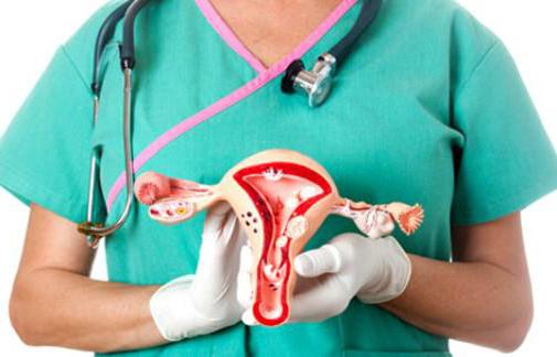
How are healthy ovaries
In order to better understand how ovarian sclerocystosis and pregnancy are related, you need to know how these organs are arranged and how they work if there is no pathology in them. The ovaries are female paired genitals. They can be represented in the form of peculiar sacs filled with brain matter. The walls of the ovaries are lined with a layer of dense connective tissue, a layer of cortical substance is located on it. It has a complex structure and importance. It is in this layer that follicles are formed - specific structural elements in which the eggs develop. Follicles, called primary, in the amount of about one to two million are laid in the body of each girl at the fetal stage. Throughout life, starting from the period of maturity and ending with the period of menopause, they are gradually consumed, and new ones are no longer formed. Therefore, the time comes when their stock runs out.
This almost never happens in women of childbearing age, so the absence of follicles can in no way be the cause of infertility. Another thing is that in their gradual ripening, malfunctions sometimes occur. So they are the culprits of the fact that the desired pregnancy does not occur. Moreover, the improper development of follicles in one hundred percent of cases leads to gynecological diseases, without treatment of which women increase the risk of thrombosis, thrombophlebitis, diabetes mellitus, heart attack, and malignant tumors in the mammary glands.
How does an ovarian cyst appear and what does it have to do with pregnancy
When the girls become sexually mature, the process of maturation of the primary follicles, which until now are as if sleeping, is included in their body. This process always goes in cycles. In each cycle, up to about 15 follicles “wake up”. They, under the action of the hormone FSH, produced by the pituitary gland, start to grow, increasing in diameter from 50 to 500 microns. During this period, a follicular fluid forms in them, and in the largest of them a cavity appears. This follicle becomes dominant, grows to 20 millimeters, protrudes. Inside it, an egg develops rapidly. The rest of the follicles from the group of “awakened” die and dissolve one after another. If everything goes according to the rules, in the female body the endocrine system is included in the work. As a result, hormones estrogen, progestins and androgens are produced, which influence the further maturation of the dominant follicle. Under the influence of the lutenizing hormone (luteotropin, lutropin, abbreviated LH), it breaks, the egg leaves it into the fallopian tube, and it turns into a yellow body and gradually dissolves.
If the rupture does not occur, the egg that has not come out is reborn, and in place of the follicle appears an ovarian cyst the size of a cherry. Those of the "awakened" follicles that did not have time to die also turn into cysts, only smaller in size. A cyst formed from a follicle sometimes grows to a considerable size (40-60 millimeters), but it may not manifest itself at all. Only in some cases, patients complain of pain in the ovary. After a woman normalizes the production of hormones, she slowly dissolves. If a woman has restored ovulation, the follicular cyst existing at that time in the ovary does not prevent the occurrence of pregnancy, but if this cyst has grown to a size of 90 millimeters, it must be removed surgically.
Causes of the disease
Scientists know in detail how ovarian sclerocystosis is formed. The reasons for this phenomenon have not yet been precisely established; there are only assumptions. Since hormones play an important role in the normal development of the follicle and the exit of the egg cell, hormonal disorders, and in particular a malfunction in the mechanism of estrogen synthesis, are considered the main cause of ovarian sclerocystosis. The following causes of hormonal disorders are called:
- heredity;
- abnormalities in the structure of genes;
- disorders in the pituitary-ovarian system;
- mental trauma;
- complications after abortion;
- infectious and gynecological diseases;
- complications after childbirth;
- changes in the functions of the adrenal cortex.
Clinical symptoms
Unfortunately, only with the onset of puberty can a girl discover ovarian sclerocystosis. Symptoms at this stage are blurry and mainly consist of menstrual irregularities. But this phenomenon can have many other causes unrelated to ovarian disease, including poor nutrition and nervous disorders. By twenty, by a maximum of twenty-five years, girls develop more specific symptoms of ovarian sclerocystosis. The main one is still a violation of the cyclicity and nature of menstruation (in 96 percent of patients). More often there are long delays in menstruation (about six months or more) or too small amounts of discharge (hypomenstrual syndrome). Much less often, patients complain about the duration and profusion of menstruation.
Other symptoms that suspect ovarian sclerocystosis are as follows:
- hirsutism (about 90 percent of patients have hairy areas around the nipples, back, abdomen, chin, and above the lip);
- overweight (70 percent of patients);
- baldness and acne on the face (found in no more than 40 percent of cases);
- some changes in body proportions;
- disturbances in the work of the nervous system;
- asthenic syndrome;
- ovarian enlargement (detected by a gynecologist on examination).
In addition, some women may experience symptoms common to many diseases: pain in the lower abdomen, malaise, inexplicably fatigue.
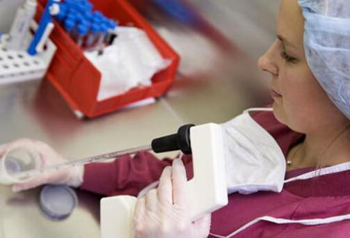
Laboratory research
Based on external signs, ovarian sclerocystosis is only suspected, and the final diagnosis is made after additional examinations. These are:
- blood test for testosterone (total should be within 1.3 ng / ml, free in women under 41 years old - within 3.18 ng / ml, and up to 59 years old - no more than 2.6 ng / ml);
- analysis of glucose susceptibility, blood sugar and triglycerides;
- colpocytogram (the material is taken from the vagina, the analysis data show whether or not ovulation is present, as well as the correspondence of the colpocytogram indices to the patient's age and phase of her menstrual cycle);
- endometrial scraping (allows you to judge about dysfunctions in the ovaries);
- control of changes in basal temperature;
- tests for some thyroid, pituitary, and ovarian hormones (LH, FSH, PSSG, prolactin, cortisol, 17-hydroxyprogesterone);
- determination of estrogen excretion.
Now patients can independently conduct a simple test that allows them to suspect cystic ovarian formations. To do this, you need a microscope (you can buy in pharmacies). In the morning, just waking up and not eating or drinking anything, you need to place a drop of your saliva on a laboratory glass and let it dry. During ovulation, estrogen levels always increase, which, in turn, changes the composition of saliva. If there is ovulation, the sample of saliva in the microscope will be in the form of fern leaves, and if there is no ovulation, in the form of dots.
Hardware Diagnostics
As a rule, for an accurate and final diagnosis, patients are prescribed in the examination complex using medical equipment.
The most gentle and completely painless method is an ultrasound diagnosis of ovarian sclerocystosis. The procedure can be transabdominal (through the stomach), transvaginal (the most informative way), transrectal (performed only in young girls and elderly women).
Ultrasound determines the size of the ovaries, their shape, structure, the number of follicles in them, the diameter of which is up to 8 mm, the presence or absence of a dominant follicle, the presence or absence of ovulation, the presence of cysts in the ovary.
Another type of examination is a gas pelvogram, showing abnormalities in the size of the ovaries and uterus.
One of the most difficult types of diagnosis is laparoscopy. It is carried out in a hospital under general anesthesia. The algorithm is as follows: the patient, the surgeon makes a puncture of the peritoneal wall and inserts a carbon dioxide pump into the peritoneum in order to create a volume in the peritoneum and to better examine the organs. Next, a laparoscope is inserted into the patient's body, which shows the status of the ovaries on the screen. Laparoscopy is the most accurate diagnostic method, but after it a woman needs a rehabilitation period.
Conservative treatments for ovarian sclerocystosis
After making the final diagnosis, in most cases, a woman is first prescribed medication. Her goal is to restore the normal menstrual cycle and resume ovulation. How to treat sclerocystic ovaries, the gynecologist decides together with the endocrinologist.
If the patient has obesity, the first stage of treatment is weight loss. A woman is prescribed a diet, feasible physical exercises.
The second step is to increase insulin perception. Metformin is prescribed, which should be taken 3-6 months.
The third stage is the stimulation of ovulation. They begin therapy with the simplest medicine, Clomiphene. The initial course is to take the drug at a dose of 50 mg overnight, starting on the 5th day of the cycle for 5 consecutive days. If there is no result (menstruation), Klomifen is taken within a month. If at the same time the effect is not obtained, the dose is increased to 150 mg per day.
The next stage (in the absence of positive dynamics) is the prescription of the Menogon medicine. It is administered intramuscularly, and at the end of the course do injections of “Horagon”. Menogon can be replaced by Menodin or Menopur.
After completing the entire course, they make blood biochemistry, and on the basis of the results of the analysis (if there is not enough LH hormone), "Utrozhestan" or "Dufaston" are prescribed.
At the same time, doctors are trying to remove excess body hair from a woman, in connection with which she is prescribed Ovosiston and Metronidazole.
A mandatory addition to the course is vitamin therapy.
Ovarian sclerocystosis: surgical treatment
If ovulation is not observed within three months after drug therapy, a woman is prescribed surgery. It is performed by several methods. Which one to apply depends on the state of the ovaries.
At the present stage, there are the following types of operations:
- cauterization of cysts using a laser;
- demedulation (removal of the middle part in the ovary);
- wedge-shaped resection (removal of the affected part in the form of a wedge from the ovary);
- decortication (the doctor removes the transformed protein layer of the ovary, pierces the follicles with a needle and sutures their edges);
- electrocauterization (point destruction in the ovary of that area in which too many hormones are produced).
- incisions (their surgeon makes a depth of 1 cm in places where the follicles are visible, so that when they mature, they can release an egg).
Forecasts
Women who agree to any methods suggested by doctors are interested in the only question: is it possible to get pregnant with sclerocystosis of the ovaries? Statistics show that without treatment, infertility is diagnosed in 90% of cases. Clomiphene drug therapy improves ovarian function in 90% of patients, but only 28% of them become pregnant. However, according to some reports, positive results can reach 80%.
The disadvantage of the drug “Clomiphene” is that it is effective only at the very beginning of the disease or after surgery as an adjuvant.
Treatment with stronger drugs, such as Gonadotropin, according to statistics, leads to ovulation in at least 28% of patients, and a maximum in 97%. At the same time, from 7 to 65% of women become pregnant.
If ovarian sclerocystosis is treated surgically, positive results are noted with approximately the same frequency as with conservative therapy. According to statistics, after surgery on the ovaries, 70-80% of women get a chance to get pregnant.
Reviews
For many women, it becomes a great misfortune to make him a diagnosis of ovarian sclerocystosis. Patient reviews about the treatment are very different. Someone was helped by pills, someone was helped by an operation, while someone did not get pregnant, despite any methods taken.
There is also a small proportion of patients reporting their pregnancy without treatment at all, although the diagnosis of ovarian sclerocystosis has not been withdrawn. Such opposite results are possible due to the individual characteristics of each person and should not be perceived as the norm.
But most women write about improving health after treatment in reviews. Only some patients report that their menstruation normalized for a short time, after which they again needed to take hormonal drugs.
And finally, there is a part of the reviews in which women note the appearance of prolonged pain in the ovaries and peritoneum after treatment with surgery.