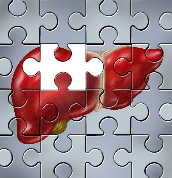
Toxic liver dystrophy is characterized by severe toxicosis, fatty degeneration and necrosis of liver cells. Mostly sucking pigs suffer from this disease along with weaners and gilts in the autumn and winter periods. At the same time, significant mortality is observed from December to January. In various industrial complexes, this pathology is observed absolutely in all periods of the year.
Etiology in animals
Toxic dystrophy of the liver develops mainly in those farms in which pigs and calves are fed biological defective feeds for a long time and kept under conditions that differ in an unsatisfactory microclimate. The main cause of toxic dystrophy of this organ is intoxication, which is caused by feeding animals spoiled and mildew-infected feeds, as well as products that contain alkaloids along with saponins and mineral poisons.
The disease is often recorded in areas characterized by a deficiency of digestible forms of selenium in soils. The cause of toxic liver dystrophy in animals can be a violation of the protein and carbohydrate ratio in the diet, along with a lack of methionine, cystine, choline, and also vitamin E. In this case, a large number of under-oxidized products are formed, which are very toxic to the body. The disease also appears with unsystematic and prolonged use of antibiotics related to the tetracycline series, and the use of other antibacterial drugs is also a factor in its development.
Toxic substances that come with food and are formed in the body against the background of digestive disorders and the metabolic process, after absorption penetrate the liver. Already in the organ, depending on the dosage and the duration of their intake, different processes appear, for example, the activity of oxidative enzymes may decrease, a sharp drop in glycogen level is noted, and at the same time fatty infiltrations develop and the breakdown of liver cells is recorded, and subsequently - liver necrosis.

Fatty infiltration in combination with organ necrosis especially progresses in the presence of a deficiency in the diet of cystine, methionine, choline and tocopherol, including. The absence or lack of these lipotropic factors leads to the fact that the newly formed fatty acid does not participate in the synthesis of phospholipids, but is deposited in the liver in the form of triglycerides. Reduction of the level of oxidation as a result of a decrease in lipolytic activity leads to the same process of fat deposition in this organ. Next, we find out what factors affect the appearance of this ailment in animals.
Causes of the disease in calves and piglets
Mostly pigs suffer from this disease along with gilts and calves during weaning and fattening. In industrial complexes that specialize in cattle fattening, on farms and in farms where they violate nutrition rules (toxic feeds are introduced in large quantities), the disease takes on a massive scale, causing significant economic damage due to forced slaughter of animals and mortality.
Toxic liver dystrophy in calves and pigs can form as a result of other diseases (infectious, gastroenteritis of various origins, sepsis), while there is an increased process of absorption of toxic products into the blood.
Symptoms
In suckling piglets, the disease most often occurs in an acute form and can be characterized by signs of arousal, rapid heart rate and intermittent breathing. Animals in this case are depressed. Body temperature sometimes rises to 40.8 degrees, and then drops and becomes subnormal. With toxic dystrophy of the liver in piglets, yellowness of the mucous membranes and skin along with vomiting is sometimes possible, and constipation is replaced by diarrhea.
Against the background of all this, a large amount of bilirubin enters the bloodstream and oliguria forms. High specific gravity urine contains bilirubin, urobilin and protein. Against the background of the occurrence of insufficiency of the heart during toxic liver dystrophy in pigs and calves, blueness of the ears, skin and abdomen is observed. This form of the disease most often leads to death.
The subacute and chronic form of the course of the disease is recorded mainly among weaned piglets, and it is characterized by less pronounced symptoms. Inhibition of general condition may be observed along with loss of appetite, sometimes vomiting and diarrhea. The temperature may be normal or low. Yellowness of the mucous membranes and skin in a chronic course is rare.
Diagnosis
The disease is determined on the basis of anamnestic information, the dynamics of the development of clinical symptoms, as a result of laboratory studies of feed, urine, blood and pathological changes in the parenchymal organs.
The anatomical change largely depends on the ethnological factor. At an early stage of the development of the disease, the liver in animals is slightly enlarged, it has a flabby texture and has a yellow color. With the development of parenchyma necrosis, flabbiness with wrinkling of the organ is more pronounced, while the shade is clay or red. A degenerative change in the heart muscle and kidneys is noted. The organ can be swollen, loosened, hyperemic, sometimes with hemorrhages, and the mucous membrane of the digestive system is covered with a viscous secret. Erosion and ulceration are observed.
Treatment
First of all, it is important to eliminate the cause of the disease. With the development of acute toxic liver dystrophy, the stomach and intestines are washed with warm water or a solution of potassium permanganate using a probe and enema. 100 grams of castor oil is introduced inside, 50 - sunflower, 30 - hemp and 100 - linseed. A hunger diet is prescribed for twelve hours. Then sick animals are fed diet food (we are talking about milk, milk, oatmeal jelly, liquid cereals from oat and barley groats, yogurt).
In addition, an acidophilic broth culture (from 20 to 40 milliliters) is prescribed twice for five to seven days. Hydrolysin is injected subcutaneously, and the lipotropic components in the form of choline chloride, tocopherol acetate and methionine are inside. Subsequently, animals are transferred to the diet from carbohydrate feed with the required amount of protein, vitamins, amino acids and mineral ingredients.
Glucose solution
At the beginning of the development of toxic liver dystrophy, subcutaneous injection of a 10% glucose solution in a dosage of 20 to 50 milliliters is effective, in addition, sodium selenite is used in an amount of 0.2 milligrams per kilogram of animal weight and intramuscular injection of calcium gluconate. The drugs are used twice a day, in the absence of glucose, you can give milk inside with sugar, hay infusion, jelly and porridge. If necessary, cardiac drugs are prescribed.
Prevention
As part of prevention, it is important to constantly monitor the nutritional value of products, the sanitary quality of feed, and at the same time the conditions of calves and pigs. According to the existing standards for each age category, the composition of the diet must include essential amino acids in the form of lysine, methionine, cystine, as well as a set of minerals and various vitamins. Any feed must be regularly examined for toxicity.
In dysfunctional farms, for prophylaxis, they recommend intramuscular administration of a 0.1% solution of sodium selenite (once twenty-five days before farrowing at the rate of 0.1 milligram per kilogram of animal weight).
Calf Dissection Protocol
According to the protocol for opening a calf, with toxic liver dystrophy in a sick animal, fatty degeneration and cell necrosis are noted. The organ, as a rule, is enlarged in volume, the capsule is unevenly colored and tense, and against the general background, foci of a yellowish color of a flabby consistency appear, which are easily destroyed by pressure. Portal lymph nodes without any changes. The gall bladder is filled with moderately thick bile, and its mucous membrane is velvety, the patency of the excretory ducts is not impaired. Based on the data of postmortem autopsy, we can conclude that the death of the animal occurred due to toxic liver dystrophy.
Pathology Features
The pathological anatomy of toxic liver dystrophy varies depending on the periods of the disease. In total, the disease takes about three weeks. In the early days, the organ is enlarged and flabby. It turns yellow. Then the capsule becomes wrinkled. Liver tissue is usually clayey. Microscopically, in the early days of the disease, fatty degeneration of hepatocytes is noted, which is quickly replaced by necrosis and autolytic decay with the formation of fat-protein detritus, in which there are tyrosine and leucine crystals.
Progressing, necrotic change captures by the end of the second week of the disease all sections of the lobules. Only at their periphery remains a narrow strip of hepatocytes. These organ changes characterize the stage of yellow dystrophy. In the third week of the disease, the liver may decrease in size and turn red. These are due to the fact that fat-protein detritus of the lobules undergoes a phagocytosis process and is resorbed.
Macrodrug
A macrodrug of toxic liver dystrophy is a glass slide on which an object prepared for research under a microscope is located. From above, this object is usually covered with a thin glass coverslip. The size of the slides is 25 by 75 millimeters, and their thickness is standardized, this greatly facilitates the storage of tools and work with them.
Conclusion
Thus, the disease involves massive progressive necrosis. As a rule, this is, first of all, an acute, much less often chronic pathological condition, which is characterized in animals by massive organ necrosis and its insufficiency. The disease develops most often with exogenous and endogenous intoxications. It is also found in viral hepatitis as an expression of its malignant form. In the pathogenesis, the hepatotoxic effect of the virus is attached importance. Allergic factors sometimes play a role.