Today, x-ray diagnostics is the most popular method for searching for disorders in the articular apparatus. Changes in bone tissues, cartilage growths, areas with a large amount of calcium deposition are only a small part of what can be detected by this diagnostic method.
Due to the ability to view the complete joint mapping on the resulting images, all problems can be identified in the early stages, as well as making the most accurate diagnosis.
What is a joint x-ray?
Before figuring out why and when an x-ray of the elbow joint is done, you first need to understand what this procedure is. In what cases is it indicated? So, x-ray diagnostics is such a way of studying the internal systems of our body. It is carried out using multiple x-rays. Thanks to the special device, the rays are sent to that area of the human body, which must be enlightened and take a picture of it.
An x-ray of the elbow joint is done according to the same principle. Rays easily pass through all soft tissues surrounding the joint, while hard tissues, on the contrary, absorb them. In this regard, in the photographs, the bones, as well as various foreign bodies, are always painted in white or light color. From this it follows that the diagnosis of pathologies associated with the skeleton with the help of such an apparatus is the most accurate. The modern world has reached the point that, for example, an x-ray of the elbow joint can be displayed not only on a special film, but also saved in a computer with the ability to display the image on the display. In this embodiment, you can increase the required area and consider it in more detail.
X-ray or MRI?
It is important to know that an x-ray of the elbow joint may not always reflect all available pathologies. This is due to the fact that there is a period when the disease first affects the soft tissues, which cannot be viewed with this device. Then other methods are included in the work: ultrasound, CT, or computed tomography, and MRI. These are also modern diagnostic methods.
MRI of the elbow joint will help to identify pathology at an earlier stage, since both soft and hard tissues will be included in the study. In the pictures you can see all the changes that occurred in the cartilage and ligamentous apparatus. Also, if you do an MRI of the elbow joint, then you can consider the nerve fibers with the vessels that feed the tissue.
In general, magnetic resonance imaging is one of the most reliable diagnostic methods in traumatology. Modern modernized devices with high accuracy help to identify pathologies in the elbow joint. MRI allows you to identify any neoplasms and the consequences of injuries.
Indications for diagnosis
If a person has constant pain in the elbow joint when bending, swelling, unpleasant crunch, then in addition to an x-ray, an MRI examination is prescribed. Moreover, an additional check is necessary in case of obtaining doubtful results of the first diagnostic method. If the elbow joint is swollen, then first of all, soft tissues are examined, since in addition to bruises, there may be suspicion of a malignant tumor.
Also, MRI is prescribed if the patient has special contraindications for examination by x-ray exposure. Often, before and after surgery, doctors require repeated magnetic resonance imaging.
What are the injuries of the elbow joint?
Injuries usually occur after a fall, when most of the body weight falls on the elbow. The results are most often fractures of varying severity, dislocations, bruises, as well as ruptures of soft tissues (muscle or ligamentous apparatus). Such injuries are common for those who have some kind of active sport in their lives. And especially for those involved in martial arts.
After such injuries, the formation of adhesions is not uncommon, which will further limit mobility. It is quite difficult to get rid of them, therefore, after an x-ray, the establishment of an accurate diagnosis and treatment, the doctor strongly recommends that you actively develop connective tissues.
The most popular injury is a dislocation of the elbow joint. The x-ray shows what exactly happened to the joint and how serious everything is. Based on the severity of the injury, doctors prescribe either conservative treatment by repositioning, or resort to surgery. Repeated x-rays of the elbow joint may be needed after rehabilitation. Also, the procedure is resorted to if the injury recurs. Moreover, in the latter case, the most often performed operation is to restore the stability of the entire elbow joint.
For fractures, conservative treatment is also used, only in the form of a cast. In case of serious injury, when the bones need to return to their previous position, endoprosthetics are performed.
In addition to elbow bruises and dislocations, patients may suffer from tendon inflammation. Many do not attach much importance to this, and pathology turns into a chronic disease, which is not so easy to cope with. In these cases, physiotherapy, joint immobilization are prescribed, and drugs are prescribed to relieve pain symptoms.
Arthrosis of the elbow
In addition to all of the above, there is such a disease as arthrosis. Often patients are delayed by treatment. Symptoms of arthrosis of the elbow joint are constant pain during flexion, extension and while walking. Sometimes discomfort can not be removed even with drugs.
If a person has a dry crunch, then this is a direct symptom of arthrosis of the elbow joint. In this case, treatment should be started immediately, since such a sound is formed due to grinding of bones against each other. Plus, there is a restriction of mobility, for example, due to muscle cramps. With advanced and chronic arthrosis, a person is usually prescribed an operation during which a damaged joint is replaced by a metal one.
Treatment of pathology boils down to the following provisions:
- It is necessary to perform exercises designed to develop a sore spot.
- Refusal to overload a sore hand.
- Use medications to reduce pain and inflammation (Diclofecan, Nise, Spazmalgon).
- Surgery indicated in the most advanced cases.
- Use of alternative treatments.
The main directions of therapy:
- Containment of pain.
- Increased mobility of the elbow joint.
- Maintaining the right lifestyle.
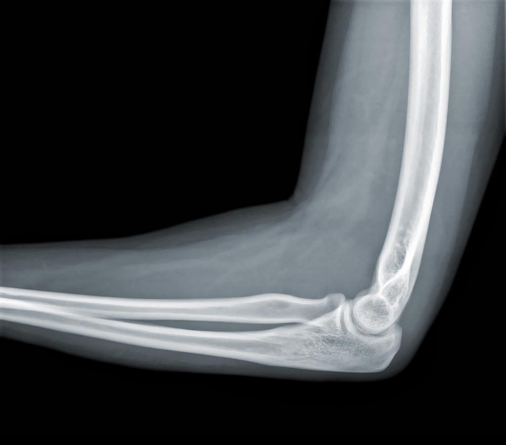
X-ray of the elbow
The elbow joint is considered the most vulnerable coupled with the patella. Since it is more susceptible to external loads. Using an X-ray of the elbow joint in two projections, it is possible to determine the nature of the damage and the structure of the bone at the end of the forearm.
The periarticular zone will also be visible in the image, which is also taken into account during the study of pathology, since often the inflammatory process can come from there.
How is x-ray performed?
In fact, there is no particular preparation for x-rays. The most important thing is to be sitting, standing or lying down for some time without movement. The average time that is allocated to the procedure is about ten minutes. X-ray examination is absolutely painless.
Before turning on the x-ray machine, the patient is covered with a lead apron in the hip area in men, the chest in women, in particular, the mammary glands. For comfort and maximum immobilization of the patient, he is placed on a table over which a special tube hangs. A photographic plate is put in the box, and the image transmitted by the device will be displayed on it.
X-ray in a hospital ward
If the patient is in the intensive care unit, then a portable version of the x-ray apparatus is used. In this embodiment, the photographic plate is placed behind the person, and the tube is mounted on a special manipulator. During the procedure, the rays are sent directly to the elbow joint and the areas closest to it.
When to do an x-ray of the elbow joint?
This part of the skeleton is a rather complex structure, including the radiopulmonary joint, shoulder-elbow and others. When should I seek the help of a radiologist:
- with any deformations or degenerative changes (a person feels discomfort, accompanied by pain);
- when edema and redness occur in the area of the injured elbow (body temperature may increase);
- with obvious and visible failures of the movable joints; with the appearance of squint.
Do not forget that any serious physical exertion can provoke serious pathology in the area of the elbow joint.
Contraindications for radiography
Basically, all existing contraindications are associated with the fact that the human body is exposed to serious radiation. But modern technology has stepped forward a long time ago, and now there are advanced models of devices that give out a lower dose of radiation than their predecessors.
However, there is still a risk for children. For example, radiation rays can inhibit the growth of a child. Therefore, age will be a contraindication: radiography can be performed if a person has reached the age of fourteen. Of course, pregnant women are exempted from the procedure. After all, radiation can damage the development of an unborn baby.
But there are exceptions when, in young children or pregnant women, severe pathologies of the elbow joints are unexpectedly detected, in which it is necessary to conduct an x-ray. In these situations, doctors try to choose the best quality device and during the examination make every effort to protect these categories of patients from radiation rays.
X-ray diagnostic options
In total, there are two types of x-ray studies: digital and analog. The first allows you to not only print the picture several times, but also display the image on the screen. It is considered the least dangerous, therefore it is used more often than analog. The second is a familiar apparatus with a film and radiation exposure.
And in that, and in another case, the doctor first installs a film or a special matrix at the level of the elbow joint in the device. After turning on the x-ray, the doctor leaves the room. To study the pathologies of the elbow joint, two or three pictures are usually taken at different angles. This is necessary for a more accurate diagnosis. Sometimes additional pictures of a healthy joint are taken, which are used in the study to compare with the damaged one.
The decryption of the result is done by the radiologist right on the spot and sends the patient to the doctor in whom he is observed. Also, the doctor can give pictures to the patient’s hands immediately. In any case, the results should be considered with your doctor.
How often can the body be exposed to such radiation?
The effect of radiation during X-ray diagnostics depends on the intensity of exposure and the time that was spent on the procedure. Of course, the effect on the body is. Irradiation is measured in doses. Each doctor has a special device in his pocket with which he monitors how many doses were received per day from x-ray studies.
For comparison, X-ray diagnosis of the colon is 6 m3t. From this we can conclude that the pictures of individual parts of the body, which are a small area for diagnosis, can be performed without much risk to health several times a year, which can not be said about comprehensive studies. Beginning in adolescence, everyone knows that fluorography is recommended to be done no more than once a year, unless the prescribed treatment requires otherwise.
With x-rays of joints, a person receives only thirty percent of the possible annual exposure. It's not that much. In turn, irradiation of the elbow joint using digital radiography is equal to three percent of the annual dose. A larger dose is always used to take pictures of bone structures than for hollow internal organs.
Of course, any method of irradiation is harmful, but the refusal of this examination can lead to serious consequences, in which the disease can go into more serious forms and stages.