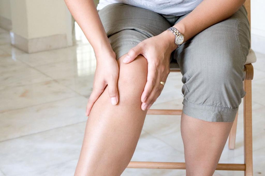
One of the serious pathologies in the body of an individual is leg vein thrombosis (photo of the disease is presented below). Blood clots formed in the vessels disrupt the natural process of blood flow. After a while, they are able to come off or completely block the venous lumen. As a result, there is a high risk of developing serious complications, such as thromboembolism, stroke, gangrene and some other equally dangerous consequences, which often cause paralysis and death of the patient.
general information
One of the biological systems responsible for the normal viability of the human body is hemostasis. Its main task is:
- blood clot dissolution;
- stop bleeding with damage to the walls of blood vessels;
- preservation of blood in a liquid state;
The formation of blood clots is one of the mechanisms of hemostasis, i.e., its appearance is necessary to stop bleeding.
A blood clot is a blood clot that formed in the lumen of a vessel when it is damaged. It covers the affected area and thus prevents bleeding. This process is called thrombosis. So, on the one hand, thrombosis protects the individual’s body from blood loss, and on the other hand, it is a provocateur of pathological conditions associated with a malfunction in the bloodstream. The causes of thrombosis:
- damage to the walls of blood vessels as a result of surgery or injury;
- change in blood flow velocity;
- high blood viscosity.
There are venous and arterial thrombosis. Next, we will talk about leg vein thrombosis (photo below). It has two forms: thrombophlebitis and deep vein thrombosis, in which the thrombus comes off and clogs the pulmonary artery.
The clot can completely cover the lumen of the vessel or only the edge of the wall. In the second case, it is called parietal and most often affects the veins of the lower extremities. Its presence provokes an inflammatory process called thrombophlebitis. Complications of thrombosis are precisely the result of untreated thrombophlebitis. Therefore, it is important not to miss the first symptoms of a blood clot.
The development of an ailment in the lower extremities. First signs
How to determine leg thrombosis at an early stage? This question is asked by many patients. At the developmental stage, a blood clot is quite difficult to detect, since during formation it is securely attached to the walls of blood vessels. As it grows, the risk of separation and the occurrence of complications is high. A small blood clot does not interfere with the nutrition of soft tissues, since it does not block the blood flow. However, even in this period there are signs indicating incipient problems with the nutrition of the soft tissues of the lower extremities. As a result, local immunity is reduced, and, as a result, the limb becomes vulnerable to the penetration of pathogenic microorganisms. Often, the fungus attacks the affected leg. An individual should pay attention to the following symptoms of leg thrombosis:
- a feeling of fullness and an increase in local temperature in the affected limb;
- seal of the dermis;
- feeling of heaviness in the legs;
- hives in caviar;
- pain when flexing the foot inward.
It is proven that early symptoms are characteristic of the female sex. The reason is that girls prefer to wear high-heeled shoes, which is a provocative factor in the emergence of various pathologies of the lower extremities. In men, the early signs of a blood clot are invisible, i.e., absent.
Late signs
Under favorable conditions, the clot increases in size and blocks the blood flow. The individual develops pronounced symptoms of leg thrombosis:
- at the location of the thrombus during palpation, a seal is felt;
- a strong pain syndrome can only be in the place of a clot or can be felt along the entire length of a blood vessel located below a blood clot;
- Intense edema gives the dermis an unhealthy appearance.
If a person does not go to a medical institution and a clot is in a vessel for a long period, then the temperature on the affected limb decreases, and the skin turns pale. Lack of treatment is fraught with the development of gangrene.
In addition, the blood clot itself serves as a provocateur of the onset of the inflammatory process. In this case, the dermis over the affected area acquires a distinct vascular pattern, becomes red and painful. The individual observed:
- Strong headache;
- increased sweating;
- general malaise;
- nausea.
Causes of Thrombosis
Under the influence of various factors in the lower extremities, the formation of clots is possible. However, the main cause of their occurrence is considered to be a malfunction in the blood coagulation system. Provocative factors are:
- insufficiently mobile lifestyle;
- violation of blood circulation;
- genetic predisposition to the appearance of blood clots;
- cardiovascular abnormalities, for example, arrhythmia, atherosclerosis;
- hormonal imbalance caused by endocrine diseases, pregnancy, as well as prolonged use of hormonal medications;
- tumor processes;
- damage to the vascular walls;
- overweight;
- bleeding disorder;
- paresis and paralysis of the legs;
- surgery, injury or injury to the lower extremities;
- septic conditions.
Regardless of the cause of the clot in the venous vessel, it must be identified and removed. Only in this case, the treatment will be effective.
Varieties
Blood clots in the lower extremities are of a different nature. Depending on the etiology, thrombosis is distinguished:
- inflammatory;
- stagnant;
- due to a malfunction of the blood coagulation system.
Depending on the location of the thrombosis is superficial and deep. In addition, the following clot forms are distinguished:
- Parietal - is considered less dangerous, since a blood clot is attached to the wall and does not interfere with blood flow.
- Occlusive - the vein is completely blocked.
- Multifocal - clots form in different places.
- Floating - in this case, the clot goes along the wall, and its top floats in the venous lumen. It comes off quite easily and getting into small vessels clogs them.
- Mixed - a blood clot moves up and down the vein.
If floating leg thrombosis is detected, what should I do? It should be remembered that this is the most dangerous form of the disease, since in case of separation, along with the blood flow, the clot easily enters the pulmonary artery. When identifying an individual, it is necessary:
- strict adherence to bed rest;
- strict observance of medical appointments, including the use of anticoagulants.
Depending on the localization of the clot, the doctor decides on the emergency hospitalization of the patient and surgical intervention.
Signs of blood clots in the lower extremities
As mentioned above, at the initial stage, the disease is quite difficult to detect. Clots form in both deep and superficial veins. An individual may suspect signs of leg thrombosis when the following clinical picture appears:
- frequent inflammation of the lymph nodes;
- temperature rise;
- trembling in the body;
- swelling;
- legs become dark blue;
- heaviness in the limbs;
- compaction in the affected area, which is clearly palpated upon palpation;
- general malaise;
- weakness, fatigue;
- the dermis is below the site of clot formation of burgundy and shiny.
In some cases, the blood clot grows slowly, and in others - quite quickly, which requires the speedy help of doctors.
If superficial veins are affected, then the symptoms of thrombosis of the vessels of the legs can appear immediately. In this case, the individual observed:
- cramps in the calf muscles;
- redness of the affected area;
- venous pattern becomes bright;
- pain during movement;
- edema;
- compaction at the site of the clot.
Symptoms of deep and superficial vein thrombosis
Usually with thrombosis, pain in the leg appears abruptly and suddenly. The limb swells, and there is a feeling that it is tearing. At the same time, body temperature rises, chills and weakness appear. External manifestations of the disease can be different, since they depend on how badly the vessel and the location of the thrombus are affected. For example, if a clot formed in the iliac or femoral vein, the entire leg swells. At the same time, blood flow worsens, and a venous network appears on the abdomen. The patient’s condition worsens dramatically. General weakness, chills, fever. Consider the differences in the symptoms of leg thrombosis (see photo in the article) for lesions of deep and superficial vessels. In the first case:
- the dermis of a diseased limb acquires a pale, and in some places a bluish appearance;
- legs increase in volume due to edema;
- a feeling of heaviness in the legs intensifies in the evening;
- an inflamed vein provokes an increase in temperature to thirty-nine degrees;
- pain in the lower leg of a bursting nature;
- the lower limb loses sensitivity, a feeling of goose bumps appears on the skin;
- pain spreads along the inner side of the thigh, lower leg and foot.
With the defeat of superficial veins:
- swelling of the foot and lower leg;
- the dermis blushes over a blood clot;
- heaviness in the lower extremities is felt;
- cramps in the calf muscles;
- goosebumps;
- pain in the leg during thrombosis along the vein increases with physical exertion;
- skin sensitivity increases.
Diagnostics
Diagnosis of lower extremity thrombosis is carried out by a doctor trained in phlebology or vascular surgery. Diagnostic Methods :
- MRI and CT - determine areas with blood clots. They are also used to identify complications.
- Impedance plethysmography - the rate of filling blood vessels with blood is determined by a change in the electrical resistance of tissues.
- X-ray phlebography is an accurate way to detect the disease. A contrast medium is injected into the vein of the lower extremities, then pictures are taken to evaluate the patency of the blood.
- Ultrasound duplex angioscanning and Doppler ultrasound are considered the gold standard for detecting thrombosis. With their help, determine the nature of the attachment of a blood clot to a vein, evaluate and analyze the degree of narrowing of the vessel, as well as the localization, type, extent and mobility of the blood clot.
- Radionuclide scanning - a radioactive substance is introduced into the vein of the foot, which can be deposited in blood clots. In the pictures, the areas affected by the thrombus are not visible.
- Blood analysis.
How to check legs for thrombosis during an outpatient visit to a doctor? To confirm the diagnosis, the following functional tests are performed:
- Mayo Pratta - initially the doctor gives the individual a massage, then imposes a tourniquet for thirty minutes. In the presence of thrombosis, pain and a feeling of fullness in the limb will appear.
- Homanza - the doctor asks the patient to lie on the couch and bend his legs. When the foot is bent, pain appears in the lower leg.
- Marching - an elastic bandage is applied over the entire leg from the fingers to the groin. After a while, it is removed, and if the patient has pain in the lower leg, then there is a chance of a blood clot.
- Lowenberg - a cuff from the tonometer is placed on the lower extremity and air is inflated with a pear, if with figures about 90 mm Hg. Art. the individual feels pain, then he has a clot in the vessel. In a healthy leg and at 150 mmHg. Art. pain is absent.
If pulmonary embolism is suspected, an X-ray of the lungs is made using a radioactive marker.
Diagnostics performed by a highly qualified specialist, as well as regular examinations, will save the individual at risk from irreversible consequences.
Methods for treating leg thrombosis
Treatment for this ailment should be comprehensive.
There are the following methods:
- Conservative includes physical activity, leg bandaging with elastic bandages, compression therapy, medication, medical nutrition, a special daily routine, the use of folk recipes, as well as minimally invasive procedures.
- Surgical.
Let's consider in more detail how to treat leg thrombosis with a conservative method. With superficial thrombophlebitis, physiotherapy is indicated, wearing compression underwear and bandaging the limb with an elastic bandage. In order to eliminate purulent or inflammatory process, the doctor recommends a course of antibacterial treatment. You can reduce the formation of blood clots with the help of special anticoagulant drugs that thin the blood:
- Heparin
- Warfarin
- "Fragmin";
- Clexane.
When they are taken, blood coagulation should be monitored by regular analysis. The dosage and duration of treatment are determined by the doctor individually.
Non-steroidal anti-inflammatory drugs used in different dosage forms will help reduce pain and inflammation: injection, ointments, gels, capsules or tablets:
- Diclofenac
- Ketoprofen;
- Voltaren;
- "Indomethacin."
Fibrinolytics are suitable for splitting a clot:
- "Urokinase";
- "Streptokinase."
Improve blood circulation:
- Trental;
- Flexital;
- Pentoxifylline.
To improve the rheological properties of blood, use:
- "Reosorbylact";
- Reftan;
- "Reopoliglyukin."
Detralex and Antistax will do an excellent job of strengthening the walls of blood vessels.
In the treatment of leg thrombosis, in order to increase the effectiveness of drug therapy, the doctor recommends the use of other methods, for example, minimally invasive procedures. If a floating clot is detected that has the ability to move around the vessel, special cava filters are installed. They are called traps in another way, they prevent the penetration of a blood clot into vital organs.
After removal of the inflammatory process, physiotherapy is possible. With contraindications to taking anticoagulants, hirudotherapy is prescribed, as a result of which the blood viscosity decreases.
Surgical treatment
And now we will consider how to treat leg thrombosis with a surgical method. It is shown with:
- ascending thrombophlebitis;
- the threat of pulmonary embolism;
- in the identification of floating clots and the inefficiency of conservative therapy;
- when a thrombus is melted with purulent contents.
To do this, use the following methods:
- Stenting.
- Thrombectomy - clot removal.
- Operation Troyanov-Trendelenburg, during which a large saphenous vein is clamped.
In particularly difficult situations, they resort to removing the affected vein. This operation is performed under general anesthesia. After the surgical intervention, the individual should begin to move as quickly as possible, i.e. walk. This is necessary to prevent the formation of repeated blood clots.
Possible complications: hidden and obvious
Untimely access to a doctor and inappropriate treatment of symptoms of leg thrombosis (see photo below) is considered to be a predisposing factor to serious consequences.
For example, an occlusal form of a clot can lead to gangrene. Fortunately, this is a fairly rare occurrence. The following complications of thrombosis are known:
- Pulmonary thromboembolism (pulmonary embolism) - blood circulation and breathing are disturbed. In the case of overlapping small branches, the patient shows signs of hemorrhagic pulmonary infarction.
- Painful blue phlegmasia - as a result of occlusion of the femoral and iliac veins, the outflow of blood is completely blocked. In this situation, there is a danger of developing gangrene.
- Purulent expansion of the clot - with thrombophlebitis in the acute stage, an abscess forms.
- Painful white phlegmasia - arterial spasm occurs next to the affected vein.
- Post-thrombotic disease - damage to the deep veins of the lower extremities with complete destruction of the valves as a result of previous thrombosis, as well as a change in the circulatory tissues.
Postpartum Thrombosis of the Lower Limbs
Often, after delivery, women are diagnosed with diseases of the venous system, including acute leg thrombosis. The reasons for this phenomenon lie in the following:
- Changes in the blood coagulation system. Both during normal childbirth and during cesarean section, there is a lot of blood loss. The body, seeking to stop it, produces several times more factors that contribute to increased coagulation. As a result, clots form in the lumen of blood vessels. In addition, during delivery, the walls of the vessels are damaged, which also provokes an increase in coagulability.
- Low tone of the vessels of the lower extremities, as well as the pelvis, is present in women after childbirth. As a result, the speed of blood movement decreases and the mechanisms of thrombosis begin to work actively.
- The hormonal background after the birth of a baby changes dramatically, which also contributes to the development of venous clots.
- Pathologies of the coagulation and cardiovascular system, overweight, anemia, late toxicosis, and also age after forty years increase the risk of thromboembolic complications.
In addition, the risk of developing this pathology is especially high in women with varicose veins.
Signs of a postpartum illness
Symptoms of leg thrombosis appear on the fifth to sixth day after delivery. With the defeat of superficial veins in the early days, there is:
- redness of the dermis along the thrombosed vessel;
- increase in local temperature over a clot;
- when touched, the vein is dense;
- deterioration in overall health;
- pain in a sore leg when moving or walking.
With the defeat of deep veins, the signs are insignificant. The main symptom in this type of leg thrombosis is edema, and it is growing and rather painful. The pain is bursting and intensifies when lowering the legs down and when standing. The dermis in the affected limb becomes milky white or blue. Basically, deep vein thrombosis is almost asymptomatic and is detected by chance with ultrasound. The danger of an undiagnosed and untreated disease in time provokes its transition to post-thrombophlebitic syndrome, in which subcutaneous tissue is destroyed, the color of the dermis changes and a fairly dense edema appears.
Treatment and prevention of postpartum thrombosis
Properly selected therapy will help to fully cope with this problem. It is important to remember that self-treatment and use of alternative medicine in the postpartum period, as well as in pregnant women, is contraindicated. If you find the above symptoms, you should immediately visit a vascular surgeon or phlebologist. What does thrombosis on the legs look like? Photos are presented in the article. An integrated approach is used in the treatment of the disease, including:
- Taking medication. The main ones are anticoagulants of the heparin group, which are approved for use by lactating women. The use of non-steroidal anti-inflammatory drugs is indicated for severe thrombosis and only after the abolition of natural feeding. Venotonics are also contraindicated in breastfeeding.
- Wearing special underwear. Compression tights, stockings or stockings help to reduce the diameter of blood vessels, normalize and increase blood flow through the veins. As a result, venous congestion and the risk of pulmonary embolism are reduced. The right doctor will help you choose the right jersey.
- Physiotherapy. In the treatment of symptoms of leg thrombosis in the postpartum period, electrophoresis, laser therapy, cryo-wraps, phonophoresis are effective. Massage, hot wraps, baths, compresses, wave types of physiotherapy are contraindicated.
In order to exclude postpartum thrombosis, preventive measures should begin during the conception planning period. If a woman in the position has varicose veins, then it will be mandatory to wear compression underwear, also a special bandage. If this ailment is detected in the second trimester, venotonics are indicated - Detralex, Antistax, Phlebodia. If a woman has a high blood coagulability, then she is recommended the introduction of injectable anticoagulants, which significantly reduce the risk of a blood clot.
Prevention
Preventive measures are especially indicated for persons who are overweight, varicose veins and lead a passive life. For the prevention of leg thrombosis should:
- rest daily by lifting the lower limbs up;
- drink at least two liters of ordinary water per day;
- do swimming or yoga;
- perform a complex of therapeutic exercises selected by the doctor;
- regularly monitor cholesterol and blood glucose;
- make a contrast shower;
- take salt baths for the legs;
- perform self-massage of the legs and feet;
- wear compression underwear;
- walk barefoot on a raised surface;
- exclude the use of oral contraceptives, as well as anticoagulants;
- do cold wiping legs;
- wear comfortable shoes, the heel should be no higher than four centimeters;
- give up smoking;
- exclude alcohol-containing drinks from the diet.
The occurrence of a clot in the lower extremities is a very dangerous condition that poses a threat to life. What thrombosis on the legs looks like is shown in the photo. Having noticed the first symptoms, you should immediately visit a doctor who, after diagnosis, will select a course of therapy.
Causes of Vein Pain
Often, pain in the legs or calf muscles is caused by inflammatory processes in the vessels. If a vein in the leg hurts, then most likely a blood clot has formed. As a result, the outflow of blood is disturbed, the walls of the vessel stretch and expand. Pain in the lower extremities is provoked by squeezing the tissues adjacent to the affected vessel. How do veins in the legs hurt during thrombosis? The individual feels a dull and pulling in nature pain. Prolonged walking and standing causes pain in the veins, as well as tension and loss of sensation. With deep vein thrombosis, pain manifests itself upon palpation. With thrombophlebitis of superficial veins, one of the patient's first complaints is pain in the vessels of the lower extremities.
Conclusion
So, leg thrombosis (photo below) is characterized by thrombosis of the veins when a clot forms in their lumen. It appears in both healthy and inflamed vessels. Most of the vessels of the lower leg are thrombosed. Blood clots form mainly in the deep veins. Most often, this disease affects the female sex due to physiological reasons.
A clot always provokes inflammation, which contributes to the formation of new blood clots. With this pathology, there is a high risk of disability or the development of complications in the form of pulmonary embolism. It is important to remember that the formation of a blood clot is a protective reaction of the individual's body. If this process were absent, the slightest bleeding would be fatal. When the vessels are healthy, the clots dissolve on their own. And in case of disruption of the coagulation and anticoagulation system, anomalies arise.