Premature retinopathy is a retinal lesion caused by insufficient maturity of the eye structure of a baby who was born prematurely. Such a pathology is characterized by visual impairment and is accompanied by clouding of the vitreous body, astigmatism, glaucoma, strabismus, retinal detachment.
The disease is diagnosed during an examination of a premature baby by an ophthalmologist using ophthalmoscopy, electroretinography, ultrasound of the eye. Treatment may include laser correction, cryocoagulation of the retina, scleroplasty.
Features of the disease
In children born much earlier than prescribed, various pathologies are almost always observed. Immediately after delivery, medical procedures and manipulations are performed that help the baby survive. However, some of them may harm him.
One of the most common pathologies is retinopathy of prematurity, which is a very serious disease of the organs of vision in a child who was born prematurely. This occurs as a result of insufficient development of the vessels of the organs of vision. Often, the disorder appears in children born before 26-32 weeks of pregnancy.
In retinopathy of prematurity, the ICD-10 code is H35.1. This pathology develops in children, even those born at the same gestational age, in different ways. In some organs of vision can fully recover, while others require surgery. In some sources, the code for retinopathy of premature infants according to the ICD is 9 (362.20), but it is almost never used. Over a period of time, the child’s body is trying to recover on its own. However, its vessels are very fragile. Their rupture leads to hemorrhage and detachment of the retina.
I must say that not all premature babies are diagnosed with retinopathy. In almost half of young patients, no abnormalities are revealed during an ophthalmological examination.
Main classification
Retinopathy of the premature is manifested in the form of damage to the organs of vision. In such a violation, three periods can be observed:
- Active.
- Cicatricial.
- Reverse development.
Active takes place until the infant is 6 months old. At the same time, the possibility of self-healing is preserved. This happens if the child develops normally, he is provided with complete peace.
The period of reverse development may be in a child aged 6 to 12 months.
Cicatricial is observed in children older than a year. In this case, the forecasts are not always positive.
In addition, many children have a malignant form of pathology. It is characterized by a very rapid development, and its distribution over the periods is not expressed enough. For an atypical form, the following types of pathology are characteristic:
- The back is aggressive.
- Pre- "plus the disease."
- "Plus the disease."
The posterior aggressive form is characterized by a very rapid transition to dangerous stages and poses the greatest threat to the newborn. It occurs quite often. The effectiveness of therapy is 50%.
Pre- “plus disease” refers to a borderline condition that manifests itself in the form of increased vascular activity.
“Plus disease” is a more accelerated form with a fairly rapid course of the active phase. It ends with retinal detachment and many other complications. With a typical course of retinopathy, the first manifestations become noticeable literally a month after the birth of the baby. Symptoms increase gradually, and the onset of the scar form occurs in about a year.
Stages of the disease
Recall that the ICD-10 code for retinopathy of prematurity is: H35.1. The disease is characterized by the presence of many violations of the organs of vision. A similar disease has several stages of development, which are rapidly growing in nature, but proceed sequentially. It is worth noting that pathology can slow down its development to any degree. The final stage of the disease can be a complete recovery or the formation of scars on the retina, provoking a deterioration in visual function or blindness.
There are several stages of premature retinopathy that have certain manifestations.
The first degree of pathology is characterized by the formation of a line between the vessels and their plane. The forecast is considered quite favorable. If for 38 weeks the pathology has not passed into an active form, then we can talk about the onset of its regression.
Retinopathy of preterm 2 stages is characterized by the formation of a roller above the demarcation line. This is considered a sufficiently alarming signal for ophthalmologists.
At the 3rd stage, connective tissue begins to form along the cushion or the tension of the vascular layer occurs and its subsequent deformation. A similar stage is characterized by the rapid formation of scars on the retina.
At the 4th stage, active retinal detachment is observed. The last 5 stage is characterized by complete detachment of the retina. In patients in this case, complete blindness is observed.
Sometimes the malignant development of the disease begins. Detachment of the retina in this case occurs without preliminary scarring against the background of the formation of swelling of the vitreous body and significant hemorrhages in the capillaries that feed the retina.
Causes of occurrence
The third trimester of pregnancy is very important for preparing for childbirth. From about 15-16 weeks, the formation of the circulatory system of the eyes begins, and the completion of this process occurs only by 36-40 weeks. In the case of premature birth, the baby has not yet fully formed the organs of vision.
The disease does not begin to progress immediately after the birth of the child, since during the first month the child’s body actively tries to independently restore the missing functions. The risk lies in the strong vulnerability of the organs of vision, which contributes to vascular damage, hemorrhage and detachment of the retina. Significantly increase the risk of retinopathy in premature babies, such factors:
- The use of oxygen in chambers for premature babies.
- Child weight up to 1500 g.
- Hereditary factor.
- Infectious diseases of the mother during the period of gestation.
- The effect of lighting on an unformed retina.
- Hemorrhages in the meninges in the fetus.
- Deviations in the development of some systems of the baby's body.
- Tobacco, alcohol and drug use by a pregnant woman.
According to statistics, most often children suffer from this disease in countries with a fairly well-developed healthcare system, which makes it possible to preserve the life of a premature baby, but their visual organs may be affected. Parents of children at risk should definitely carefully monitor their child’s health and consult an ophthalmologist in a timely manner, even if there are no visible signs of a disorder.
Signs and Symptoms
The visible signs of premature retinopathy do not appear immediately, since it is very difficult to detect visual impairment in a newborn baby. All children at risk are surely given the advice of an ophthalmologist. Parents may notice some characteristic symptoms on their own. Among them, you need to highlight the following:
- During the game, the child brings toys close to his face.
- Observes everything that happens with only one eye.
- Does not see objects located far away.
- Does not respond to external stimuli.
- Blinks with only one eye.
- The baby has a pronounced squint.
- A sharp decrease in vision.
If scarring occurred during retinopathy of premature infants, there are also five degrees of this pathology:
First one. Vision is not impaired, since the fundus remained unchanged.
The second one. The center of the retina has shifted, the peripheral areas are changed, which in the future increases the risk of retinal detachment.
The third. The center of the retina is offset significantly. The area where the nerve enters is deformed in it.
Fourth. Creases are clearly visible on the retina, which significantly impairs vision.
Fifth. Retinal disinsertion.
Diagnostics
Diagnosis of retinopathy of prematurity is carried out at the age of 3-4 weeks. You can determine the onset of the disease using the following methods:
- Ophthalmologist examination with an ophthalmoscope.
- Digital retinoscopy.
- Ultrasound diagnostics.
- Electroretinography.
- Optical tomography.
In addition, you need to pass a general and biochemical blood test, a bacterial analysis of a conjunctival smear, and an ECG. If a child has a plus disease, an examination should be carried out every 3 days. In the presence of the first two stages, the eyes of the child are examined every week to confirm the regression of the disease. In the future, preventive examinations by an ophthalmologist are required up to adulthood, and at an older age according to the doctor’s testimony.
Fundus examination is carried out using special means (dilating pupil and soft eyelids) to exclude unnecessary, traumatic effects on the child’s eyes. Before the onset of stage 3 of the disease, confirmation of its regression is necessary. Otherwise, surgery is recommended to preserve vision.
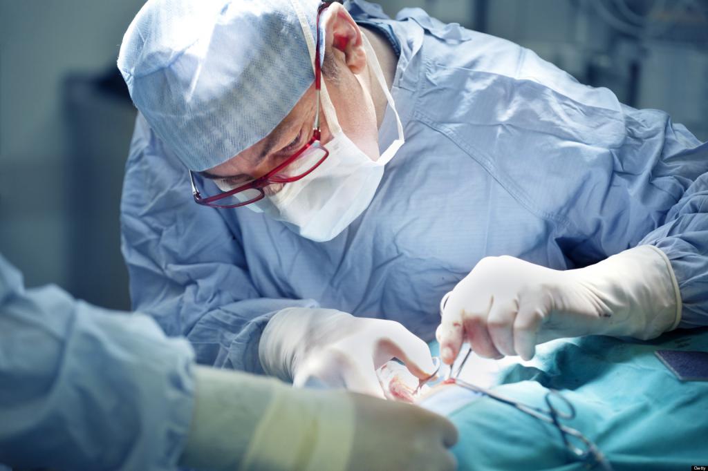
Features of surgical treatment
Parents whose children are at risk need to know exactly what retinopathy is premature and how to treat the disease. You need to trust doctors and understand that in the first 2 stages, special therapy is not required. The child only needs regular monitoring by an ophthalmologist. Often the disease independently regresses.
Treatment can be carried out in 2 ways:
Conservative therapy is rarely used. Often the child is sent for surgery. At the 3 stages of the disease, laser or cryosurgical retinal coagulation is used. In this case, a complete cessation of the formation of scar tissue is observed and the disease regresses.
Basically, the procedure is performed under general anesthesia, which may be associated with a possible risk of deterioration of the heart muscle. That is why ophthalmologists prefer to carry out laser coagulation, since this is a completely painless method with a minimum of side effects. When conducting such an intervention, a scar forms in the problem area, which prevents the growth of vascular tissue. With this method, regression can be achieved even with the occurrence of complicated forms of retinopathy.
If the disease has passed into the cicatricial stage, then scleroplasty is performed. This is a mechanical combination of the retina with the detachment area. In the case of a successful operation, vision is significantly improved. If such a technique was ineffective, then a vitrectomy is performed, during which the scar tissue, and in some cases the lens of the eye, is removed. Sometimes the second stage of the operation, as well as the subsequent implementation of laser coagulation, may be required.
With retinopathy, premature surgery is effective only in the first year. If you hold it later, it is unlikely to help increase visual acuity.
Conservative therapy
At this time, there is no single approach to the treatment of retinopathy of premature infants, since this process depends on many different conditions. Some doctors say that surgery is carried out only at the 3 stages of the pathology, and the first two require constant monitoring of the patient and conservative therapy. Others insist on an earlier operation.
A conservative technique is suitable for the treatment of the early stages of the disease. In order to maintain the health of the child, the following groups of medications are used:
- Hormonal drops ("Prenacid", "Maxidex", "Dexamethasone").
- Antioxidants (Emoxipin, Ascorbic Acid).
- Angioprotectors ("Etomzilat", "Ditsinon").
For the diagnosis and treatment of retinopathy of premature infants, clinical recommendations have been approved and approved by the Joint Commission on the Quality of Medical Services of the Ministry of Health of the Russian Federation. The methods noted in the protocol were considered in this article.
During conservative treatment, preparations containing vitamin E are also used. They have an antioxidant effect, which together with other drugs helps to achieve a positive result.
During the period of regression of the disease or during recovery after surgery, physiotherapy may be prescribed. In this case, apply the following procedures:
- Electrophoresis
- Magnetostimulation - speeds up the recovery process.
- Electrical stimulation - normalizes metabolic processes and improves blood supply to the organs of vision.
Possible complications
Even after successful completion of the operation, the consequences of retinopathy of premature infants can be quite dangerous, there is a high probability of complications. The risk of negative manifestations is very high with untimely therapy or after a severe course of the disease. Among the main consequences it is necessary to highlight the following:
- Dystrophy of the eye.
- Cataract.
- Glaucoma.
- Retinal disinsertion.
- Myopathy
- Amblyopia.
- Astigmatism.
That is why, after treatment, the child should always be under the supervision of specialists who will conduct regular examinations. If the child does not comply with the established standards of visual acuity, especially after treatment, he is assigned disability.
Prevention and prognosis
To prevent retinopathy, efforts must be made even during pregnancy planning. If the baby was born ahead of schedule, then you need to conduct regular examinations of his organs of vision.
If there is a need for oxygen therapy, it is necessary to control the level of oxygen. At stages 1-2 of retinopathy, the prognosis is positive, since the disease goes away on its own. There is a high probability of recovery in 3 stages, if appropriate treatment has been carried out.
Reviews
On the treatment of retinopathy of premature parental reviews, in general, favorable. Many mothers are alarmed by the requirement to conduct examinations once a week. But those parents who have already gone through this report that there is nothing to worry about visiting a doctor every week.
Some mothers report that their children retinopathy went away without any treatment, but they were observed by an ophthalmologist until the diagnosis was made.
Those parents whose children survived the operation also write in the reviews that there is nothing scary in it. Kids after surgical treatment spend a day in a hospital, where they are monitored. After they are discharged home. If the disease is detected in time and all the requirements of doctors are met, eyesight in children with retinopathy is fully restored.