A complex disease in which stones sink into the ureter is always accompanied by severe pain. This pathology is dangerous for its complications if adequate treatment is not undertaken in a timely manner. Doctors, calling such an ailment ureterolithiasis, give him the second highest prevalence in urological practice. Pathology can be detected in children. Most often, it is diagnosed in men. But sometimes stones are found in the fair sex in the ureter. Symptoms in women usually indicate a severe course of the disease.
Pathology characteristic
Urolithiasis is a fairly common disease. Its appearance provokes many different factors. Most often, pathology occurs against the background of poor nutrition and unsatisfactory quality of drinking water. Initially, stones are formed in the kidneys.
Most patients for a long period of time do not even suspect the presence of calculi. After all, the symptoms of the disease do not appear immediately. Meanwhile, calculi "grow" in the kidneys. And as a result of certain factors, stones may appear in the ureter.
What are the symptoms in women? This is, first and foremost, a great pain. It indicates renal colic (indicates a lowering of the calculus in the ureter). In such situations, you should immediately consult a doctor.
Penetration of the stone into the ureter
Calculi, as a rule, are formed in the renal pelvis. However, there are cases when stones were formed in the ureter. Symptoms in women, treatment - these are moments that must be discussed with a doctor. Self-control of pathology is completely unacceptable.
So, if calculus forms in the kidneys, why does it get into the ureter? Provoke such a movement can many different factors. Most often this occurs as a result of the following reasons:
- carrying weights;
- long shaking trip;
- plenty of fluids and food;
- horseback riding.
It is very important to remember what signs appear if stones in the ureter are localized. Symptoms in women, indicating the progression of calculus, are manifested in the form of severe pain. Acute discomfort appears in the stomach and back. This condition is called renal colic.
Causes of the disease
Ureter calculi are formed from various substances:
- uric acid;
- cystine;
- calcium phosphate ;
- struvite.
Most often, the following factors influence the process of stone formation:
- Genetic predisposition. Doctors say that more often the disease is diagnosed in patients who have cases of urolithiasis in the family.
- Disturbed outflow, stagnation of urine. Congenital pathologies may underlie the development of the disease. Most often the disease is provoked by narrowed ureters in women, their underdevelopment, kinks or anomalies of the bladder.
- Diseases of the urinary tract in a chronic form. Diseases of an infectious nature can lead to the development of pathology. For example, pyelonephritis.
- Broken exchange. Acquired or congenital ailments may be accompanied by the penetration into the urine of lithogenic substances - calcium (if hyperparathyroidism is diagnosed), urate (in case of gout).
- Digestive system diseases. If the suction function is impaired, stones may form.
- The use of drugs. Some medications can lead to the development of the disease. For example, such effects provoke uroseptics from the category of nitrofurans.
Doctors say that uroliths often form in women living in hot and dry climates. High-calorie foods rich in animal proteins are capable of starting the mechanism of the development of the disease.
Symptoms of the disease
There are times when stones in the ureter do not cause severe pain. Symptoms in women that characterize the movement of the calculus are completely dependent on its size and shape. Stones not exceeding 2 mm in diameter are able to move painlessly along the ureter. In this case, no symptoms may be observed. A woman will not even know about an unpleasant pathology in the body.
But most often there are large stones in the ureter in women. Signs of pathology provokes a stuck calculus.
In this case, the symptoms are pronounced and are called renal colic:
- Sharp, severe pain localized in the lumbar region. She gives women in the perineum and labia.
- Urination may be impaired. But such a sign is observed extremely rarely and characterizes the simultaneous exit of stones from both ureters. Most often, women have a frequent urge to urinate.
- In the urine, there is blood and an internal epithelium of the kidney. Such symptoms appear as a result of damage to the ureter with sharp edges of the calculus. If the stone completely blocked the path, then there will be no such sign, since urine enters only through the normal ureter, not affected by the disease.
- Hanging sweating, chills. There is an increase in temperature to 37–37.5 degrees. Pathology can be accompanied by nausea, flatulence, often vomiting.
The calculus usually progresses periodically. This leads to the fact that the painful symptoms in a woman either appear or disappear. Such colic can annoy for several hours or days.
Symptoms of pathology, depending on the location of the calculus
Most often, the calculus is found at the site of narrowing of the ureter. This is the area in which the renal pelvis connects to the canal. This area is called the pyelourethral segment. The next area in which a stuck stone is often diagnosed is the area where the ureter transitions from the pelvis to the pelvis. Another “dangerous” area is the connection of the canal with the bladder.
If the calculus clogs the ureter in the upper zone in women, the symptoms are as follows:
- severe pain appears in the lower back;
- acute discomfort is wavy in nature, now subsiding, then intensifying;
- a change in body position does not reduce the intensity of pain;
- discomfort covers the lateral abdomen.
The following signs indicate the localization of the stone in the middle zone of the channel:
- pain is acutely felt in the lateral abdomen (below, along the edge of the ribs);
- discomfort extends to the inguinal and iliac regions.
If the calculus descended into the lower part of the ureter, then the woman's symptoms appear as follows:
- the pain is localized in the lower abdomen and groin;
- extreme discomfort covers the labia majora;
- urination becomes frequent;
- there is a feeling of fullness of the bubble;
- the process of urination does not bring relief (the feeling of emptying does not appear).
Possible complications
It is very dangerous if there are stones in the ureter for a long time. Symptoms in women, treatment of pathology require a serious and responsible attitude.
Otherwise, severe consequences may develop, such as:
- hydronephrosis;
- acute renal failure;
- fistulas on the ureter;
- obstructive pyelonephritis.
Diagnostic Methods
To make sure that severe discomfort is triggered by the movement of calculus along the ureter, the doctor will conduct an initial examination. It involves palpation.
Then the patient will be assigned more accurate studies:
- urinalysis, which determines protein, salt, pus, blood cells;
- back sowing;
- analysis of urine to study its acidity;
- X-ray examination;
- blood analysis;
- urography;
- Ultrasound of the urinary tract;
- CT scan of the kidneys;
- radioisotope diagnostics.
The complex of such examinations allows you to determine the location of the calculus, identify the sources of the disease and select the appropriate therapy.
Treatment methods
If during the diagnosis, stones in the ureter of women are revealed, only a competent specialist can decide how to remove them.
The treatment methods depend on the complexity of the situation, the size of the calculus. Depending on these factors, they can develop in 2 directions:
- Conservative-waiting therapy. It is undertaken in cases where the stone in diameter does not exceed 2-3 mm and does not clog the duct. In this case, the possibility of an independent exit of calculus is high.
- Active treatment. Used if conservative therapy is not possible or has not yielded positive results.
Drug treatment
How to remove a stone from the ureter?
Conservative-waiting therapy includes:
- The appointment of urolitic drugs. Medications "Nifedipine" or "Tamsulosin" provide acceleration of the passage of calculi.
- The use of painkillers, antispasmodics. Often, the patient is recommended NSAIDs, such as Ibuprofen, Naproxen.
- The woman is prescribed physiotherapy and special physiotherapy exercises.
In addition, the doctor recommends that the patient review her diet.
Dieting
Of particular benefit will be dietetics. It is based on the exclusion of foods from the diet that contribute to the formation of stones in the body, and recommends increasing the consumption of food, accelerating the withdrawal and dissolution of calculi.
To provide such recommendations, you must:
- Refuse food containing oxalic acid (cabbage, spinach, nuts, currants, legumes).
- Do not combine the above foods with dairy products that are rich in calcium.
- Include foods containing vitamin A (broccoli, carrots, pumpkin) in your diet.
- Every week, arrange a fasting day (watermelon or cucumber).
- Establish a drinking regimen. Drink about 2 liters of water daily.
Why is specialist help needed?
Sometimes the above conservative therapy is ineffective, and stones in the ureter are still diagnosed. Symptoms in women, removal of calculus is important to discuss with a professional urologist. It is strictly forbidden to fight the disease on your own.
Self-treatment can lead to rather sad consequences. Among these complications, urinary tract infection often appears. And this is a direct path to the development of sepsis. Unfortunately, in advanced situations, the patient may even be assigned to remove the ureter, and sometimes the kidney.
Surgical intervention
Most often, to eliminate the calculus, which is stuck in the ureter, the following methods are used:
- Lithotripsy. The most effective way to crush stones. Moreover, it is less traumatic. Lithotripsy involves the distant crushing of stones using waves. The event lasts about 1 hour on average. It is carried out in most cases without anesthesia.
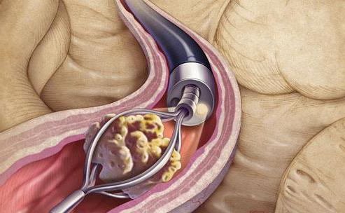
- Urethroscopy This removal of calculus is carried out using a special device introduced into the canal through the urogenital system. Sometimes, before the introduction of the urethroscope, the stones are first crushed by a laser. Intervention is carried out under general or partial anesthesia.
- Ureterolithotomy. This is a surgical intervention, which is justified with fairly large stones. During this operation, the calculus is removed through the dissection of the walls of the ureter. Of course, the procedure involves general anesthesia.
Stones in the ureter are a serious pathology in which it is extremely dangerous to procrastinate on a visit to the hospital. The disease refers to severe ailments, which can lead to disastrous results. Therefore, do not practice self-disposal of stones. Seek help from competent professionals.