Modern medicine makes a lot of efforts for the prevention or timely detection of various pathologies of the mammary glands. But, despite the abundance of information, there are many that remain beyond the interests of a wide audience. Microcalcinates in the mammary gland - what is it and why are they dangerous? What causes this phenomenon and how to deal with them? Let's try to figure it out.
Microcalcifications in the mammary gland - what is it?
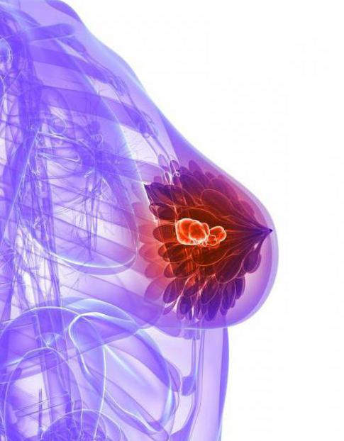
A phenomenon such as micro-calcifications, or calcifications (deposits of calcium salts), is quite common. They appear on the site of dead or irreversibly altered tissues in various human organs. Most often, this becomes the result of some kind of inflammatory process. Similar formations can be observed in pathological processes in the lungs, kidneys, liver, prostate. The thyroid gland and heart do not avoid the same fate. In this case, there may be signs of the underlying disease, as well as the absence of any noticeable changes in well-being. The accumulation of microcalcifications in the mammary gland can signal a serious illness such as breast cancer. Fortunately, the identification of these formations does not mean an unconditional sentence - oncology is confirmed only in 30% of cases, and the remaining manifestations relate to benign changes. Nevertheless, if even a single microcalcinate was detected in the mammary gland, this is an occasion to undergo a serious examination.
Reasons for the formation
In addition to the oncological pathology of the mammary glands, many processes in a woman's body can provoke the formation of microcalcifications. The most common of these are the following:
However, in addition to physiological reasons, these formations are also present with a number of pathologies of the mammary glands. With sclerosing adenoses, fibrocystic mastopathies , microcalcifications in the mammary gland are also found. What it is? These pathologies are considered benign processes. Most often, they are accompanied by pain that intensifies during menstruation. A cyst in the mammary gland can be manifested by pains of a rather intense nature, extending to the axillary region, shoulder or shoulder blade. To the touch with these pathologies, it is possible to determine the areas of compaction, depending on the form of the disease, having clear boundaries or passing in the form of cords and fine grain.
Symptoms with microcalcifications
The danger of microcalcifications in the mammary glands lies in the fact that their growth process is completely asymptomatic. If a painful seal in the mammary gland is easy to detect and take action in time, then calcifications do not show anything due to their small size. They do not cause pain, discomfort or fever. It is possible to detect them only with an X-ray examination. In the picture, microcalcifications in the mammary gland are visible as blackouts having a certain shape and localization. It is precisely by these parameters that the mammologist determines the presence of a pathology and diagnoses it.
Classification
As a rule, microcalcifications in the mammary gland alone are not the cause of any pathological processes. But their presence often becomes an important diagnostic factor, indicating a certain pathology. In order to make it easier to establish an accurate diagnosis, they are usually classified according to several parameters.
According to the localization (location) of calcifications in the mammary gland, there are:
By the nature of their distribution:
segmental - calcifications are located in one lobule of the mammary gland;
regional - accumulation of calcifications is located within one share;
linear - salt deposits are visually arranged in a line;
grouped - the accumulation volume does not exceed 2 centimeters;
diffuse - single calcifications are randomly distributed over the chest.
In addition, calcifications are divided according to their shape:
Stromal microcalcifications
Calcinates of this localization are most safe from the point of view of the diagnosis of cancer pathologies. The place of their accumulation are the walls of blood vessels, fibroadenomas, skin or fatty cyst in the mammary gland. The cause of their formation is most often necrosis of adipose tissue and fibrous formations. As a rule, stromal calcifications differ in relatively large sizes, but do not have clear boundaries. If scattered microcalcinates in the mammary gland are formed in the space of the sebaceous glands, then they have a clear oval or close to rectangular shape. All this makes it easy to identify them during x-ray examination.
Lobular calcifications
Lobular calcifications are most often due to atrophic changes in the glandular tissue. Moreover, they have a rather characteristic appearance - clearly defined rounded formations located within one or more lobes. As a rule, the presence of such deposits almost always indicates the presence of tumor processes. In 80% of cases, especially if during examination a painful compaction in the mammary gland is revealed, and on one of the proportions of the x-ray, shapeless spots resemble crescents or bowls, it is possible to diagnose fibrocystic mastopathy with confidence. However, to exclude oncology, a biopsy is additionally prescribed.
The highest probability of a malignant process can be with heterogeneous cotton-like or powder-like clusters. It is believed that the finer the calcifications (their size can vary from 50 to 500 microns), the more heterogeneous their shape and random location, the greater the likelihood of detecting oncology.
Duct calcifications
Ductal calcifications are formed in the ducts of the mammary glands. If they are formed due to diseases such as mastitis or ectasia of the ducts, they have clear worm-shaped contours, interrupted structure and localization, which coincides with the duct. It is also possible the formation of calcifications in the form of points or small segments with blurry, vague contours. This is highly likely to indicate a malignant process.
Diagnostics
A mammologist is engaged in the diagnosis of this pathology. The problem is that with palpation of the breast, it is impossible to detect not only scattered microcalcifications in the mammary gland, but also quite large formations. You can see them only on an x-ray. That is why it is so important for women older than 40 years to do a mammogram at least once a year. This study allows you to identify the appearance of microcalcifications in time, and it is not difficult for an experienced specialist to determine which disease caused their appearance. In some cases, when such formations are detected, in order to exclude breast cancer, a biopsy of breast tissue is additionally performed. In addition, a biochemical blood test is mandatory and a hormonal background is examined.
Treatment and prevention
In the event that microcalcifications were found in the mammary gland, treatment will depend on what caused them to appear. If a histological examination of the collected material confirms the presence of a malignant tumor, then oncologists will do this, and depending on the severity of the process, chemotherapy will be performed or surgical treatment will be prescribed. In the event that the process is benign, a mammologist can prescribe hormone therapy, breast massage, and a corrective diet.

As for prevention, it largely depends on the woman’s ability to take good care of her health. We all do not live in the most favorable environmental conditions, often we eat not the most wholesome foods and lead not the most healthy way of life. But it’s enough to single out one day a year, visit a mammologist, take the necessary tests and do a mammogram to know for sure microcalcifications in the mammary gland - what it is, whether you have them or not, and what causes their appearance. And if the reason for the appearance of calcifications is serious enough, then timely diagnosis will help to take the necessary measures in the early stages of the disease.