Basal pneumosclerosis is a disease that is accompanied by severe irreversible changes in lung tissue. The lesion occurs in the area of the respiratory organ where the main bronchus is located, as well as the pulmonary arteries, veins and nerve nodes. With this pathology, healthy lung tissue (parenchyma) is replaced by connective tissue. In the affected areas, gas exchange is sharply disrupted, which leads to severe shortness of breath and hypoxia. Over time, dangerous complications come from the heart, mediastinum, and blood vessels.
Causes
Basal pneumosclerosis always occurs due to other bronchopulmonary pathologies:
- obstruction of the bronchi and lungs;
- pneumonia and bronchitis of an infectious etiology;
- lung injuries;
- inflammation of the pulmonary vesicles (alveolitis);
- pleurisy;
- tuberculosis
- bronchiectatic disease (expansion of the bronchi with suppuration);
- Beck sarcoidosis (benign pathology of the lymphatic vessels);
- exposure to respiratory organs of allergens, smoke, polluted air, toxic substances;
- casting (reflux) from the stomach into the airways of undigested food.
This disease often occurs after chronic inflammatory processes occurring with frequent exacerbations, as well as after untreated pneumonia or bronchitis.
Provocative factors
Many diseases can cause pneumosclerosis. However, this complication does not develop in all people who have undergone bronchopulmonary pathologies. The following factors can provoke a sclerotic process in the lungs against the background of diseases:
- decreased immunity;
- exposure to radiation (including radiation therapy);
- frequent colds;
- hypothermia.
In addition, congestion in the pulmonary vessels, as well as impaired lymph outflow, can cause dystrophic changes. Therefore, basal pneumosclerosis is often formed in patients with heart failure of the right ventricle and pulmonary vein thrombosis.
Pathogenesis
Sclerotic changes in the lungs arise as a result of a prolonged inflammatory process. The vessels lose their elasticity, and the basal areas become dense and stiff. Normal lung tissue is replaced by fibrous tissue.
Pulmonary vesicles (alveoli) in the affected areas stick together. As a result, the lungs lose their shape and decrease in size. Sclerosed zones lose their functions and cannot support gas exchange.
Subsequently, the fibers of the pathological tissue penetrate into the vessels of the lungs and nerve plexuses. Pathological changes sometimes develop rapidly, and the affected areas do not have time to overgrow with connective tissue and become covered by cysts.
Dystrophic changes can affect any part of the lung. It is necessary to distinguish between basal and basal pneumosclerosis. In the second case, pathological changes occur not in the region of the main bronchus, but on the periphery. The basal form is characterized by slow development. Substitution of the parenchyma with connective tissue in the basal zones is much more active than in the peripheral regions.
Due to serious gas exchange disorders, a constant oxygen deficiency is formed in the body. This affects the condition of many organs, and in the first place - the heart and brain.
Is pneumosclerosis contagious
Often, patients ask the question: "Is basal pneumosclerosis contagious or not?" This disease cannot be transmitted from one person to another, as it has a non-infectious etiology. The patient is absolutely not dangerous to others. Pneumosclerosis in the basal regions occurs as a serious complication of bronchopulmonary diseases, including infectious ones (for example, pneumonia or tuberculosis). But this pathology itself is not contagious.
Forms of the disease
Pulmonologists distinguish local and diffuse basal pneumosclerosis. What are these diseases? What is the difference?
With local pneumosclerosis in a small area of the lungs, seals occur. The gas exchange function is not impaired, and the patient does not have any complaints. The disease is detected by chance during the examination.
With the diffuse form of pneumosclerosis, anatomical changes in the structure of the lung tissue occur. Large areas of connective tissue form and their number increases over time. The lungs decrease in volume, and their vessels also undergo pathological changes.
There are several degrees of connective tissue distribution:
- Fibrosis. Separate sections of fibrous tissue form.
- Sclerosis. Connective tissue gradually replaces healthy tissue, changes in blood vessels occur.
- Cirrhosis. Collagen grows in blood vessels, bronchi and alveoli. There is a severe gas exchange disorder. This is the most difficult stage of the disease.
Symptomatology
Basal pneumosclerosis of an initial nature proceeds without clearly expressed symptoms. Periodically, a person feels increased fatigue. Most often, this is attributed to hard work or age-related changes. With large physical exertion, shortness of breath appears. In most cases, patients do not pay attention to the early signs of developing hypoxia.
In the future, it becomes difficult for a person to breathe even with little physical exertion. He is concerned about shortness of breath while climbing stairs and when walking fast. Respiratory difficulties progress as more and more lung zones are involved in the pathological process.
In the later stages of the disease, the patient experiences shortness of breath even while walking at a slow pace or during a conversation. Due to developing hypoxia, a person feels constant fatigue and weakness.
In the basal part of the lungs there are a large number of nerve nodes and plexuses. Therefore, sclerosis of this site is accompanied by chest pain. At first, the discomfort is aching. Over time, the pain becomes paroxysmal and intense.
Short-term respiratory arrest may occur periodically. This symptom greatly scares the sick. Over time, a person is able to take only shallow breaths. The patient develops signs of respiratory failure:
- bluish-pale skin tone;
- tachycardia;
- headache;
- chronic fatigue;
- prostration;
- insomnia.
In the later stages, a person experiences episodes of loss of consciousness, swelling of the face and limbs occurs, and heart failure forms.
Patients are coughing. At the beginning of the disease, it occurs only in the morning, and by the middle of the day passes. As the pathology develops, the cough becomes constant and painful, with sputum difficult to separate.
"Pulmonary heart" is a symptom of a late stage of basal pulmonary pneumosclerosis. What does this mean? Due to obstructive and dystrophic changes in the respiratory system, pressure in the pulmonary circulation increases. According to this system, blood flows from the right atrium into the right ventricle, then passes through the pulmonary vessels and returns to the left atrium. Hypertension in a small circle occurs due to changes in the arteries and veins of the lungs. As a result, the patient’s right ventricle greatly increases. "Pulmonary heart" is manifested by the following symptoms:
- severe shortness of breath even with complete rest;
- swelling on the face and limbs;
- pain in the sternum;
- swelling of venous vessels on the neck;
- sensation of pulsation in the lower abdomen;
- low body temperature.
Such serious pathological changes in the heart often lead the patient to disability.
The symptomatology of the disease also depends on the localization of the sclerotic process. Crepitus while listening to the lungs is a sign of lower lobe radical pneumosclerosis. What it is? During auscultation, you can hear a characteristic crisp sound when you inhale. It occurs due to the adhesion of the affected alveoli.
With the defeat of the upper lobes, the appearance of the syndrome of "drumsticks" and "watch glasses" is possible, it is observed in the late stages of basal pneumosclerosis of the lungs. What is this syndrome? The fingers of the patients thicken in the region of the terminal phalanges and become like drumsticks. Nails also undergo deformations, their plates become thick and convex, which resembles watch glasses.
The mechanism of deformation of the fingers and nails is not fully understood. It is assumed that due to hypoxia, the vessels of the terminal phalanges expand, which leads to the proliferation of tissues.
Diagnostics
The doctor begins a diagnostic examination with an examination and analysis of the patient's complaints. In this case, it is very important to identify a history of bronchopulmonary diseases. It is such pathologies that most often cause pneumosclerosis. Auscultation of the lungs is mandatory. Above the affected area, weakened breathing, wheezing, and sometimes crispy sounds are heard.
After a preliminary examination, the doctor prescribes an X-ray of the lungs. This is the most reliable research method for pneumosclerosis. An x-ray shows the localization and prevalence of dystrophic changes.
On the radiograph, the affected area looks like a blackout with uneven edges. The sclerosed part is reduced compared to the same area of a healthy lung. The vessels and pleura are pulled to the affected area.
Connective tissue normally should not be visible on an x-ray. With pneumosclerosis, fibrotic changes look like a mesh-like pattern.
To study respiratory function, spirography or pneumotachography is prescribed. The patient breathes in a special tube, and the instruments fix the volume of exhaled air, the vital capacity of the lungs and the speed of inspiration and expiration.
Drug treatment
How to treat radical pneumosclerosis? Dystrophic changes in the lungs are irreversible, and it is no longer possible to restore the normal function of the affected areas. You can only try to stop the spread of sclerosis and conduct symptomatic therapy.
With the local form of pneumosclerosis, only dynamic observation is indicated. Drugs are not prescribed, however, the patient must periodically undergo an X-ray of the lungs and visit a doctor.
In the symptomatic treatment of basal pneumosclerosis in diffuse form, the following groups of drugs are used:
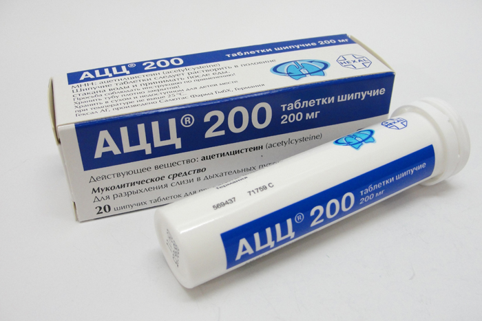
- Mucolytics (“ACC”, “Mukaltin”, “Lazolvan”, “Erespal”). These are expectorants that thin the mucus and help remove it from the bronchi.
- Antispasmodics ("Theofedrine", "Isadrin", "Fenoterol"). Such drugs relieve spasms of the bronchi and facilitate breathing.
- Antibacterial drugs and sulfonamides. These drugs are indicated if pneumosclerosis has developed against the background of an infectious pathology.
- Corticosteroid hormones (Prednisone, Dexamethasone, Hydrocortisone). These drugs are prescribed in the form of injections or inhalations in severe cases of the disease.
- Non-steroidal anti-inflammatory drugs ("Nise", "Diclofenac". "Ibuprofen"). They are prescribed for severe chest pain.
- Cardiac medications (Asparkam, Adoniside, Strofantin, Digoxin). With pathological changes in the lungs, an increased load on the myocardium is created. These drugs help maintain the functionality of the heart muscle.
- The drug "Cuprenil." This agent inhibits collagen production and reduces the proliferation of connective tissue.

Pneumosclerosis is a serious illness. It is often accompanied by weight loss and weakness. Therefore, multivitamin complexes must be included in the complex therapy.
If necessary, drug therapy is supplemented with bronchoscopy. Using this procedure, the respiratory tract is drained and drugs are delivered directly to the bronchi.
Oxygen therapy
Oxygen therapy plays an important role in the treatment of this pathology. The procedure saturates the patient's body with moist oxygen and prevents hypoxia. Gas is supplied using a special apparatus in the following ways:
- Through the mask. This method is suitable for patients who are in satisfactory condition and can breathe on their own.
- Through a catheter. In this case, O 2 is supplied through a tube inserted into the nasal passage. The method is used if the patient requires a constant supply of oxygen.
- Intubation way. This method is used if the patient is in serious condition. Humidified oxygen is supplied through a tube inserted into the patient's trachea.
- In the pressure chamber. In the absence of consciousness, the patient is placed in a special chamber and oxygen is supplied into it under a certain pressure.
Oxygen therapy gives good results in patients with radical pneumosclerosis. The patients' reviews indicate that after a course of procedures, they showed signs of hypoxia and significantly improved well-being.
Physiotherapy
With pneumosclerosis, physiotherapeutic treatment is widely used. The following procedures are prescribed:
- electrophoresis with iodine preparations;
- inductothermy and diathermy on the chest area;
- iontophoresis with calcium chloride and novocaine;
- ultraviolet radiation.
If the disease is in a compensated stage, then physiotherapy exercises are indicated. However, in this case, you need to strictly dose physical activity and follow the advice of a doctor. Useful breathing exercises. They are best carried out in the fresh air, gradually increasing the duration and intensity of classes. If the patient has a fever or cough with blood, then breathing exercises are strictly prohibited.
Doctors also recommend therapeutic chest massage sessions. This helps reduce blood stasis and suspend the sclerosis process.
Cell therapy
Currently, research is underway in the treatment of radical pneumosclerosis. A method of treatment using stem cells is currently considered the most promising.
Stem cells are administered intravenously. With blood flow, they enter the affected area and replace damaged cells. As a result, normal tissue is restored in the lungs and gas exchange is normalized.
How effective is this treatment? The success of cell therapy depends on the stage of the disease. Treatment should be started as early as possible before fibrotic changes affect most of the lung. After all, a supply of healthy tissue is needed in order to start the recovery process.
Surgery
Surgical treatments for basal pneumosclerosis are rarely used. The operation is indicated only with the local form of the disease, if the compaction in the lungs suppurates or there is tissue destruction. In this case, the affected area is removed.
Forecast
What is the prognosis of radical pneumosclerosis of the lungs? Life expectancy depends on the severity of the pathology. Mild forms of the disease are treatable. The sclerotic process can be slowed down and suspended. Severe forms that occur with complications can lead to death from heart and respiratory failure.
The local form of pneumosclerosis is usually asymptomatic and has a favorable outcome. In most cases, the disease does not progress, and compaction in the lungs is limited to only one small focus.
A more complex prognosis is diffuse basal pneumosclerosis of the lungs. Life expectancy with such a pathology will depend on the effectiveness of the prescribed treatment. It is important to remember that sclerotic changes in the lungs always develop against the background of other pathologies. The outcome of pneumosclerosis largely depends on the severity of the underlying disease.
Prevention
How to prevent radical pneumosclerosis? First of all, it is necessary to cure bronchopulmonary diseases on time and to the end. It is also important to strengthen your immunity through hardening and physical activity. It is necessary to stop smoking, because even passive inhalation of tobacco smoke can lead to lung pathology.
Every person needs to do lung fluorography regularly. People working in hazardous industries need to undergo special preventive examinations. This will help to identify pulmonary pathologies at an early stage and prevent irreversible changes in the respiratory system.