A defect in the interventricular septum in children is a congenital anomaly that occurs during the period of intrauterine development of organs, the heart in particular. This defect, along with other congenital heart defects, is most common in medical practice. Statistics in numbers reaches 42% among all congenital heart defects. Moreover, the gender factor does not play any role: equally often both boys and girls suffer from the disease. However, a direct genetic relationship from the closest blood relatives has been proven. Doctor of Medical Sciences Belozerov Yu. M. in his work “Children's Cardiology” indicated that the likelihood of developing a defect in this case increases by 3.3%.
Congenital defect of the interventricular septum of the heart is characterized by impaired blood flow in the large and / or small circles of blood circulation. Along with all existing malformations, heart disease remains the main and most common cause of early mortality in children.
For a better understanding of the essence and characteristics of this defect, it is necessary to go a little deeper into epidemiology.
Epidemiology
The interventricular septum is involved in the process of contraction and relaxation of the heart muscles. In the fetus, it is fully formed at 4-5 weeks of development. With any deviations in the development process, a defect remains in it, which subsequently violates hemodynamics and causes further health problems. In 80% of cases, a defect is detected along the perimeter of the membranes. In the remaining 20%, there are defects in muscle genesis.
Defect classification
A ventricular septal defect is a congenital heart defect. Typically, these defects are divided into large, small and medium. To assess its size, resort to comparison with the diameter of the aorta. Defects from 1 to 3 mm in size are small, they are located in the muscle wall of the interventricular septum, and are called Tolochinov-Roger disease. It is characterized by good visualization during diagnosis, as well as a minimum of hemodynamic deviations and, by and large, in itself does not pose a danger to the human body. In this case, the aforementioned defect can be detected during a random examination, this is a kind of physiological feature that may not manifest itself in any way throughout a person’s life, and all that may be required is just control in dynamics. Another thing is large multiple defects, measuring 1 cm or more, with obvious vivid symptoms that threaten health and life. In this case, doctors strongly recommend that the operation be performed in the first 3 months of the child’s life in order to minimize possible complications and death.
Departments of the interventricular septum
The interventricular septum consists of three sections: the upper (its densest part) is membranous, adjacent to the connective tissue, the middle part strengthens the heart muscle, and the lower trabecular part forms a spongy structure.
Based on the anatomical location, defects are divided as follows:
- the perimembranous defect of the interventricular septum is 75% of all defects, located in the upper part under the aortic valve; may spontaneously close;
- muscle defects of the septum - up to 10% of interventricular defects, are located in muscle tissue, remotely from valves and conductive systems;
- epigastric defects - make up the remaining 5%, are located above the supraventricular ridge, and are unable to close spontaneously.
Hemodynamics
What happens to a baby born with a ventricular septal defect? How does a defect manifest itself?
Intracardiac disorders begin to form not immediately from the moment of birth, but on 3-5 days. In such an early period, heart murmurs may not be heard at all, due to the same pressure in the ventricles due to pulmonary hypertension. Later, when the pressure in the pulmonary artery gradually decreases, there is a fragmentation of pressure in the ventricles - this leads to the release of blood from the high-pressure area to the low, from left to right. This leads to the fact that the right ventricle experiences a constant supersaturation of blood, an additional volume overflows the vessels of the pulmonary circulation - this is how pulmonary hypertension develops.
Stages of Pulmonary Hypertension
It is customary to distinguish the stages indicated by the Russian cardiac surgeon Burakovsky, a specialist in the field of congenital heart defects in young children.
- Hypervolemic stage. It all starts with stagnation of blood and is aggravated by pulmonary edema, decreased immunity and, as a result, frequent infections, but most importantly, the development of severe pneumonia and poorly treatable. If there is no response of the body to conservative treatment, they resort to surgery - narrowing of the pulmonary artery according to Mueller. The operation allows you to artificially narrow the lumen of the artery, which for some time will provide a smaller discharge of blood into the pulmonary circulation. However, the effect of this method is not long-term, and reoperation is usually required after 3-6 months.
- Over time, the Kitaev reflex triggers in the vessels. The bottom line is that the vessels react to overload and stretching with spasms. And this is followed by an imaginary hidden course of the disease, a transitional stage. At this time, the child can stop getting sick, he becomes active, gaining weight. This condition is most favorable for the operation.
- High pulmonary hypertension - Eisenmenger syndrome. In the long term (in the absence of surgical intervention), vascular sclerosis develops. This is a very dangerous and irreversible process. At this stage, cardiac surgeons most often refuse to undergo surgery for patients, due to the lack of reasonable guarantees for the cure and the severity of the patient's condition. When listening, 2 tone above the pulmonary artery is pronounced, systolic murmur becomes weak or it is completely absent. They fix the appearance of Graham-Still noise, diastolic murmur due to valve insufficiency. The characteristic signs are wheezing and hard breathing, the chest protrudes in the form of a dome and is called the “heart hump”, cyanosis is aggravated - the skin and mucous membranes become cyanotic, and from peripheral cyanosis develops into diffuse.
Causes of ventricular septal defect
Let us turn to the causes of this defect.
Most often, a defect in the interventricular septum in children is formed at the stage of laying the organs and is caused by intrauterine developmental disorders. This defect is often accompanied by other cardio-disorders. From 25 to 50% of cases, the defect goes side by side with abnormal development of the kidneys, mitral valve insufficiency and Down syndrome. A direct negative effect during embryonic development is caused by endocrine disorders in the pregnant body, viral infections. The negative impact of the environment and bad ecology, the effects of radiation, the use of alcohol and drugs, as well as the termination of pregnancy and toxicosis, can affect. Not the last role is played by hereditary factors. It is worth noting that the main reason for the development of the acquired defect is complications after myocardial infarction.
Clinical picture
The clinical picture with defects of the interventricular septum consists of a whole complex of symptoms characteristic of heart failure. As a rule, they develop by 1-3 months of a child’s life. It all depends on the size of the above defect. The occurrence of early protracted bronchitis and pneumonia indicates the presence of a defect. Upon closer inspection, the child looks pale and lethargic, inactive, drooping, with a characteristic lack of vital interest in the gaze. Shortness of breath can be detected, which is noted both during physical activity and at rest, tachycardia, expansion of the heart borders or their displacement. A symptom called "cat purr" is also characteristic, stagnant wheezing is heard. As a rule, systolic murmur is very intense, goes to the right side of the sternum, is heard in the IV intercostal space to the left of the chest and from the back. A pathological increase in the size of the liver and spleen during palpation is noted. In children, hypertrophy develops very quickly.

Diagnostic Methods
The standard diagnosis of any heart disease consists of an X-ray examination of the chest organs, ultrasound, ECG - electrocardiography, and two-dimensional Doppler echocardiography, MRI. X-ray examination gives a superficial description of the shape of the heart, determines the size of the cardiothoracic index.
An ECG will show if there is an overload of the ventricles, and later of the atria, the heart, and hypertrophy - all of them are witnesses of a high degree of pulmonary hypertension.
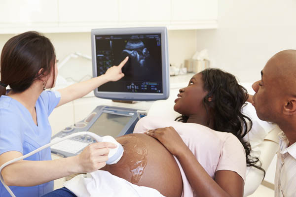
What will the ultrasound show?
Ultrasound with a defect of the interventricular septum in the fetus allows you to identify many heart pathologies at the very early stages of development, it is a simple widely available, and at the same time, informative research method.
The study provides the following heart information:
- the size of the cameras, their structure and integrity;
- the condition of the walls of the heart, the presence or absence of blood clots and neoplasms in them;
- the amount of fluid in the pericardial sac;
- pericardial condition;
- the thickness of the walls of the chambers of the heart;
- condition and diameter of coronary vessels;
- valve structure and functionality;
- myocardial behavior at the time of contraction and its relaxation;
- blood volume during its movement in the heart;
- possible noises in the organ;
- the presence of infectious lesions;
- defects of partitions.
Doppler echocardiography
Doppler echocardiography clarifies the exact location of the defect, its size, as well as the pressure in the right ventricle of the heart and pulmonary artery. For the first stage of pulmonary hypertension, indicators up to 30 mm Hg are characteristic, for the second stage, indicators range from 30 to 70 mm Hg. Art. In the third - the pressure is above 70 mm.
Magnetic resonance imaging
MRI examination is the most informative and indicative method of examination. It visualizes the current state of tissues and blood vessels, helps to establish or clarify the diagnosis, suggest the subsequent development of the disease and develop an optimal and appropriate treatment plan. Timely diagnosis of a defect in the interventricular septum undeniably increases the chances of preventing the aggravation of the disease and the speedy recovery of the patient.
Treatment
Treatment of heart disease with a defect in the interventricular septum consists of conservative treatment and surgical intervention in heart tissue. Conservative therapy includes drug therapy. As a rule, we are talking about inotropic drugs in combination with diuretics. In the case of surgery, a palliative surgery is performed - narrowing of the lumen of the pulmonary artery according to Muller. Or radical defect correction is carried out - a patch is made from pericardial tissue.
Forecast
There are frequent cases when defective “holes” by themselves close by the end of the mother’s pregnancy, or some time after birth. This happens because, after birth, the baby’s circulatory system starts up again, and the pressure changes - all this can positively affect the baby and lead to healing. A doctor can only prescribe supporting medications, and regular ultrasound.
If there is a defect in the interventricular septum, one should not remain inactive and let the course of the disease take its course. Only in extremely rare cases does a defect have no significant effect on quality and life expectancy.
For the most part, it poses a real danger and threat to human life. Life expectancy with the aforementioned defect directly depends on the size of the defect, but on average the numbers vary from 20 to 25 years. More frightening reality indicates statistics from 50 to 80% of child mortality before reaching 6 months or one year of age. Therefore, it is very important to recognize the first signs and symptoms, to identify the disease in time and to take the right and timely steps to eliminate it.