Aortic heart defects usually have an acquired character and are clinically manifested only in old age. Their presence can cause severe hemodynamic disturbances. The severity of the pathology is that the changes affecting the valves are irreversible.
Heart structure: valves
The heart is a hollow organ that consists of 4 chambers. The left and right halves are separated by partitions, in which there are no formations, however, between the atrium and the ventricle of each side there is an opening equipped with a valve. These formations allow you to regulate blood circulation, preventing regurgitation, that is, reverse casting.
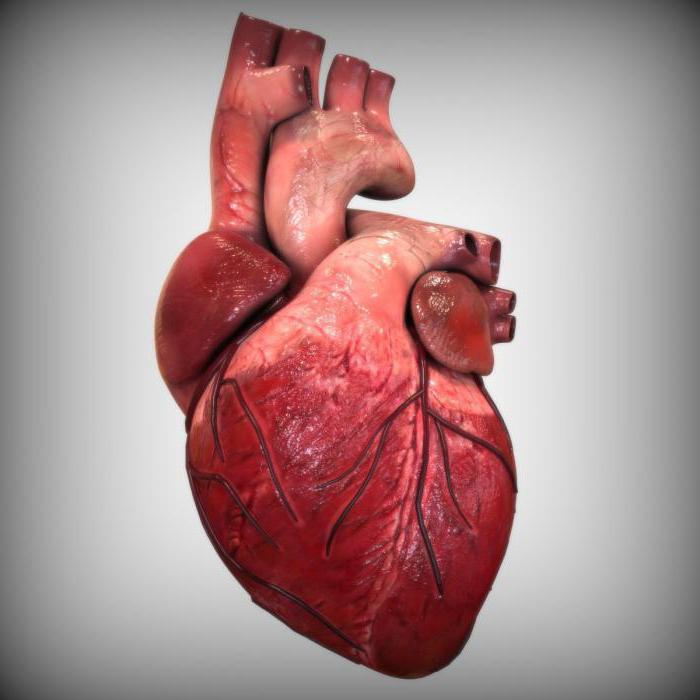
On the left there is a mitral valve, consisting of two valves, and on the right there is a tricuspid valve , it has three valves. The valves are equipped with tendon threads, which ensures their opening only in one direction. This prevents backflow of blood in the atria. At the junction of the left ventricle into the aorta there is an aortic valve. Its function is to provide unilateral advancement of blood into the aorta. There is also a pulmonary valve on the right side . Both formations were called "lunate", they have three wings. Any pathology, for example, calcification of the valves of the aortic valve, leads to a disruption in the movement of blood. Acquired defects are usually associated with a disease. Therefore, people with so-called risk factors should undergo regular examinations: mainly an echocardiogram.
Aortic valve mechanism
Aortic valve plays a significant role in blood circulation. Shutters are sealed or shortened - this is one of the main pathologies. It becomes a cause of hemodynamic disturbance. The function of this part of the organ is to ensure the movement of blood from the left atrium into the ventricle, preventing regurgitation. The valves are open during the period of atrial systole, at which time the blood is directed through the aortic valve into the ventricle. Further, the shutters are closed to prevent back casting.
Heart defects: classification
By the time of occurrence, it is possible to distinguish congenital heart defects (aortic valve and other formations) and acquired. Changes affect not only the valves, but also the septum of the heart. Congenital pathologies are often combined, which makes diagnosis and treatment difficult.
Aortic valve stenosis
Pathology implies a narrowing of the transition of the left ventricle to the aorta - valve flaps and surrounding tissues are affected. This disease, according to statistical indicators, is more common in men. Compaction of the walls of the aorta and the valves of the aortic valve is usually associated with rheumatic and degenerative lesions. Also, the role of the etiological factor can be endocarditis, rheumatoid arthritis. These diseases lead to fusion of the cusps, as a result of which their mobility decreases, and the valve cannot fully open during the systole of the left ventricle. In old age, the cause of the lesion is often atherosclerosis and calcification of the valves of the aortic valve.
As a result of narrowing of the aortic orifice, significant hemodynamic changes occur. They are observed when stenosis has a pronounced degree - a decrease in the tract by more than 50%. This leads to the fact that the pressure gradient of the aortic valve changes - in the aorta, the pressure remains normal, and in the left ventricle it increases. An increased effect on the wall of the left ventricle leads to the development of compensatory hypertrophy, that is, to its thickening. Subsequently, diastolic function is also disturbed, which is the reason for the increase in pressure in the left atrium. Hypertrophy leads to an increase in oxygen demand, however, the increased blood supply falls on the increased mass of the myocardium, and even with reduced pathologies. This leads to the development of heart failure.
Clinic
In the early stages, the affected aortic valve may not manifest itself in any way. Clinical changes occur when the opening is narrowed by 2/3 of the norm. With severe physical exertion, patients begin to worry about pains localized behind the sternum. In rare cases, pain can be combined with loss of consciousness due to systemic vasodilation. The formation of pulmonary hypertension leads to shortness of breath, which at first only bothers with exercise, but then appears at rest. The protracted course of the disease causes chronic heart failure. Pathology requires surgical treatment, as there is a risk of worsening and sudden cardiac death.
Diagnostics
On examination, patients have a characteristic pallor associated with reduced cardiac output. The pulse on the radial arteries is difficult to feel - it is rare and weak. With auscultation, a weakening of 2 tones or its splitting is observed. The ECG is not sufficiently informative - signs of hypertrophy are determined only with a severe degree of stenosis. The most indicative echocardiography, allowing to evaluate the aortic valve. The valves are sealed and thickened, the opening is narrowed - these are the main diagnostic criteria that this study helps to detect. The degree of stenosis and the pressure gradient effectively determine the catheterization of the cavities.
Treatment
With mild to moderate stenosis, only lifestyle correction is required - avoidance of excessive physical exertion, treatment of concomitant pathologies. With an increased frequency of contractions, adrenergic blockers are prescribed, and with heart failure, diuretics and cardiac glycosides are effective . Severe compaction of the walls of the aorta and the valves of the aortic valve requires surgical treatment. As a rule, prosthetics or balloon dilatation are performed .
Aortic valve insufficiency
This name was given to a pathology characterized by non-closure of the valves. This phenomenon leads to a reverse reflux of blood into the left ventricle, which occurs during the diastole. Defect is usually a complication of infectious endocarditis and rheumatic fever. Less commonly, syphilis, aortic aneurysm, aortitis, arterial hypertension, Marfan syndrome, systemic lupus erythematosus lead to it.
The aortic valve plays an important role in blood circulation. Incomplete closure of its valves becomes the cause of regurgitation, that is, the return of blood into the left ventricle. As a result, an excess volume of blood arises in its cavity, which leads to overload and stretching. Systolic function is impaired, and increased pressure leads to the development of hypertrophy. The pressure in a small circle increases retrograde - pulmonary hypertension forms.
Clinic
As with stenosis, pathology makes itself felt only with a pronounced degree of insufficiency. During exercise, shortness of breath occurs, it is associated with pulmonary hypertension. Pain bothers only in 20% of cases. At the same time, auscultatory and external manifestations of the pathology are expressed:
- Ripple of the carotid arteries.
- Symptom Durozier or the occurrence of systolic murmur on the femoral artery. It occurs when it is pinched closer to the listening position.
- Quincke's symptom is a change in the color of the lips and nails in accordance with the pulsation of the arterioles.
- The double tones of Traube are loud, “cannon” that arise over the femoral artery.
- Symptom de Musset, manifested by shaking his head.
- Diastolic murmur after 2 tones arising from auscultation of the heart, as well as a weakening of 1 tone.
Diagnostics
Informative methods are echocardiography and catheterization of cavities. They allow you to evaluate the aortic valve, as well as to track the volume of regurgitated blood. Based on these studies, the severity of the defect is determined and the question of the need for surgical intervention is resolved.
Treatment
Severe insufficiency with a large volume of regurgitation, intense clinical manifestations requires surgical treatment. The optimal output is artificial aortic valves, which allow you to restore heart function. If necessary, symptomatic drug therapy is prescribed.
Stenosis and aortic valve insufficiency are the most common heart defects, which, as a rule, are the result of any local or systemic disease. Pathology progresses quite slowly, which allows you to diagnose it in time. Modern methods of treatment help to restore the valve and improve the patient's condition.