Tetralogy of Fallot is a congenital heart disease, which is characterized by several abnormalities at once. In the presence of such a disease, the functioning of the cardiovascular system changes, which is fraught with more dangerous consequences. For example, in newborns, this pathology is most often accompanied by cyanosis, shortness of breath and severe developmental delay. That is why parents are so worried about what the disease is, under the influence of what factors it develops, what symptoms are accompanied. Indeed, early diagnosis and appropriate treatment is the only thing that can provide a child with a normal life in the future.
What is tetralogy of Fallot?
Tetralogy of Fallot (ICD) is a congenital, complex heart disease. The morphological basis of the pathology consists of four signs (which, in fact, is evidenced by the term "tetrad"). It:
- right ventricular outlet obstruction;
- aortic displacement;
- hypertrophy of the right ventricle ;
- extensive ventricular septal defect.
This pathology was first described in 1888 by the French pathologist Fallot, whose name was subsequently named. According to statistics, this disease accounts for about 7-10% of congenital malformations. By the way, sometimes tetralogy of Fallot is combined with other anomalies, for example, open ductus arteriosus, stenosis of the branches of the pulmonary artery, right aortic arch, etc.
The main causes of the development of the disease
Tetralogy of Fallot is a pathology that is formed as a result of a violation of the bookmark process and the development of the cardiovascular system at about 2-8 weeks of embryonic development. It is worth saying that the causes of this anomaly are not fully understood. On the other hand, the researchers managed to find out some risk factors.
For example, a pathology can occur if a woman suffered an infectious disease (rubella, measles, scarlet fever and some others) in the early stages of pregnancy. Violation of the bookmark and development of the fetal organs may be associated with the use of certain drugs, including hormonal drugs, sleeping pills and sedatives. Taking drugs, alcohol also affects the formation of congenital heart disease (CHD).
Tetralogy of Fallot is sometimes diagnosed in children with other anomalies, for example, the so-called Amsterdam dwarfism. This is a serious disease, which is accompanied by many disorders, including deformation of the auricles, hypertrichosis, myopia, atrophy of the optic nerves, spinal deformity, etc.
As a rule, the development of this heart disease begins from the moment when, for one reason or another, the arterial cone rotates counterclockwise. As a result of improper rotation, the aortic valve is displaced, and the aorta itself is located on the interventricular septum. As the fetus develops, the pulmonary trunk lengthens, narrows and shifts due to improper aortic positioning.
Disease classification
Tetralogy of Fallot in children can be represented by four types, depending on obstruction of the excretory tract of the right ventricle:
- Embryological, or the first type of Fallot's tetrad, is a form of pathology in which the conical septum is displaced to the left and forward. The fibrinous ring is not deformed, and the maximum zone of stenosis is located in the muscle ring.
- The second type, or hypertrophic Fallot tetrad, is a kind of pathology in which there is not only a displacement of the septum (as described in the previous case), but also its pronounced hypertrophy. The zone of maximum development of stenosis is observed at the level of the output section of the right ventricle.
- The third type of pathology, also called tubular in medicine, is characterized by an incorrectly formed division of the common arterial trunk. As a result of such a violation of the normal structure, a change in the pulmonary cone is observed - it becomes narrowed and shortened. Also, for this type of disease, stenosis of the pulmonary artery and hypoplasia of the fibrinous ring are characteristic.
- A multicomponent, fourth form of tetralogy of Fallot, as a rule, is accompanied by a significant lengthening of the conical septum.
Features of hemodynamics in this disease
What changes are accompanied by heart disease? In any case, the Fallot tetrad entails hemodynamic disturbances, the severity of which is determined by the form of obstruction of the excretory duct of the right ventricle, as well as by the presence of changes in the interventricular septum.
If stenosis of the pulmonary artery is significant and there is a large septal defect, then a larger volume of blood from both ventricles enters the aorta. But much less than normal is supplied to the pulmonary artery, which provokes the development of arterial hypoxemia. In the extreme form of this pathology, blood enters the pulmonary circle of blood circulation from the aorta either through the collateral or through the open arterial duct.
If the patient’s obstruction is moderate, then a left-right blood discharge develops - in this case, an acyanotic form of the disease is observed. Nevertheless, the disease is constantly progressing - at first the blood collection becomes cross, and then right-left, while the disease is transformed from a white form to a blue (cyanotic) one.
What are the symptoms of the disease?
The clinical picture with this pathology can be different, since it largely depends on the form of the disease. For example, sometimes a tetralogy of Fallot in a newborn is accompanied by severe cyanosis. In this case, the baby may suffer from shortness of breath during feeding. Also in the first months of life there is a low increase in body weight. On the other hand, if the stenosis of the pulmonary artery in a child is minimal, then at rest, cyanosis is not observed, and therefore it is not always possible to diagnose the disease by external signs quickly.
Quite often, hypoxia attacks develop against the background of motor activity. This condition is accompanied by deep and very frequent breaths. The kid begins to scream, becomes extremely restless. Cyanosis is gradually increasing. As a rule, this picture is observed in young children, and the symptoms are most pronounced at the age of two to four months.
If tetralogy of Fallot in a newborn is accompanied by severe attacks, this can lead to general lethargy and convulsive seizures. Sometimes pathology leads to death. Older children sometimes instinctively squat during a physically active game, which presumably reduces systemic venous return and increases oxygen saturation in arterial blood.
The main symptoms of the pathology are attacks of cyanosis and shortness of breath, which are often accompanied by loss of consciousness. The degree of oxygen deficiency and the severity of attacks directly depend on the severity of pulmonary stenosis.
It is also worth noting that in the clinical picture, as a rule, one can distinguish several stages of the relative well-being of the child, as well as several especially difficult periods:
- From birth to 6 months. The fact is that during this period, children are not too physically active, and therefore cyanosis and hypoxia are moderate. Naturally, this does not always happen, some children suffer almost from the first days of life.
- The most difficult stage (from a medical point of view) is observed from six months to two years. The attack develops with a spasm of the excretory section of the right ventricle. As a result of a spasm, all venous blood is discharged into the aorta, which leads to the development of more pronounced hypoxia of the brain. As a rule, an attack in a child begins suddenly - it becomes restless. Cyanosis and shortness of breath are observed, sometimes up to a loss of consciousness. Convulsions often appear. This process in most cases occurs against a background of emotional or physical stress.
In the future, the clinical picture changes slightly. At an older age, cyanosis is constantly growing (sometimes quickly, sometimes slowly), but attacks almost completely disappear. Shortness of breath and tachycardia are reduced, and a collateral circulation pattern is formed in the lungs.
Modern diagnostic methods
Naturally, only a qualified doctor can make a similar diagnosis. Tetralogy of Fallot is a pathology that requires a complete medical history and a series of studies that will not only confirm the presence of a congenital malformation, but also help determine the form of the disease, its severity, and the general state of the child’s health.
In the presence of symptoms, such studies are carried out:
- The initial examination includes an electrocardiogram. During the procedure, signs of overload of some parts of the heart, as well as hypertrophy, can be noted. For example, results may demonstrate weak sinus syndrome or AV blockade.
- Tetralogy of Fallot is a pathology that can be noticed during an X-ray examination of the chest. For example, this defect is accompanied by bulging of the arc of the left and right ventricles, there is a noticeable depletion of the pulmonary pattern. In newborn children, the heart on the radiograph in shape often resembles a shoe.
- A rather informative method is echocardiography. During this study, you can accurately assess the size of the cavity of the right ventricle, as well as determine the type of pathology, evaluate the degree of narrowing of the outlet duct. Using the same method, heart valves, in particular, the aortic valve, are examined, the general condition of its valves is examined, and the diameter of the fibrous rings is measured. The fact is that if aortic stenosis is not detected and aortic treatment is performed, complications, in particular, pulmonary edema, may occur at the end of therapy.
As a rule, the studies described above are enough to determine if a child has a defect. Tetralogy of Fallot, however, is a diagnosis that is sometimes very difficult to make, and a number of additional procedures may be required.
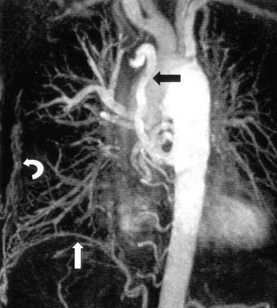
For example, coronary angiography is sometimes performed. This procedure is necessary in order to determine the exact location of the coronary and main vessels, to check for the presence of any concomitant anomalies, and also to accurately determine the type of heart disease.
Catheterization is a more complex procedure, which includes puncture of the heart cavities. This study is prescribed if you need to measure pressure in the right ventricle, to determine the degree of blood oxygenation in the aorta.
Tetrad Fallot: treatment
The treatment regimen largely depends on the form of the disease and its severity. Medication helps to eliminate seizures and prevent the development of some complications associated with oxygen starvation.
As a rule, infants are prescribed infusions of prostaglandin. This procedure sometimes helps to open the arterial duct again. If the baby shows the first signs of an attack, it is recommended to press his knees to his chest. Older children squat themselves - this position helps to avoid the appearance of a hypoxemic attack.
Intravenous fluid administration is recommended to supplement the volume of circulating blood. In some cases, small patients are prescribed morphine. If necessary, blood pressure is increased with phenylephrine or ketamine (this substance, by the way, also has a sedative effect). Propranolol sometimes helps prevent relapses.
Sometimes oxygen therapy is performed to stop the attacks, but it is worth noting that the effect of such treatment is quite moderate.
Palliative Surgery
Naturally, various drugs can alleviate the condition of the child. Nevertheless, drugs can not save the patient from such a dangerous ailment as tetralogy of Fallot. Surgery today is the only way to eliminate the defect. Only surgical intervention is able to give the baby a chance at a normal growing up and a full life.
Today, there are two main methods of surgical treatment. Palliative surgery involves the imposition of various types of inter-arterial nodes, which allows you to restore blood circulation. For example, the most popular procedure today is the application of subclavian pulmonary anastomosis. During the procedure, the subclavian artery is connected to a unilateral pulmonary artery using a special implant.
Radical surgery as a treatment method
Doctors recommend that if possible immediately carry out a radical operation, as it gives much better results. As a rule, such a procedure is carried out from the age of 6 months to 3 years - it is during this period that it is most effective (if we consider the further process of growing up and developing the child).
A radical method of treatment is the complete correction of pathologically altered structures. With the help of a patch, surgeons close the ventricular septal defect, expand the stenotic area of the pulmonary artery. Naturally, the procedure is complicated, and therefore only a qualified heart surgeon can carry it out.
Tetralogy of Fallot: prognosis for patients
Naturally, first of all, parents are interested in the prospects for such a disease. Tetralogy of Fallot in children - the pathology is extremely serious, it can in no way be ignored.
It should be understood that the course of the disease largely depends on the severity of pulmonary stenosis. If we are talking about statistics, then, unfortunately, almost a quarter of the children die in the first year of life. Without surgery, the average age of children with an ailment is 12 years. Only about 5% survive to 40 years old with a diagnosis such as tetralogy of Fallot. Disability, developmental problems, constant monitoring and poor quality of life - this is what, unfortunately, adult patients have to face.
An alternative, of course, is available. Do not forget about surgical treatment, which is often prescribed for children with a diagnosis of tetralogy of Fallot. After the operation, children, as a rule, recover quickly, develop normally, even are able to withstand large physical exertion. A pattern has been noted - surgery at a later age does not give such good results in the future as an operation performed in infancy.
Patients with this pathology, even after the end of therapy, should undergo regular examinations by a cardiac surgeon and cardiologist. Before any surgical or dental procedures, patients should undergo special treatment to avoid the risk of developing endocarditis. Naturally, there are other requirements, limitations in the patient’s life, which the attending physician will definitely tell about.