Sacralization of the L5 vertebra is a congenital pathology that is characterized by full or partial fusion of the last lumbar vertebra with the sacrum. Of course, people who are faced with a similar problem are looking for any additional information.
What is a pathology? What are the reasons for its development? What symptoms should I look for? Is complication possible? What treatment methods can a doctor offer? The answers to these questions interest many people.
Anatomical features
Normally, between the fifth lumbar (L5) and the first sacral (S1) vertebrae, there is the strongest and most stable intervertebral disc. It performs a number of important functions:
- reduces the effect on the spine during walking and movements (plays the role of a shock absorber);
- supports the necessary dimensions of the intervertebral region, since here important nerve fibers that innervate the lower limbs and pelvic organs depart from the spinal cord;
- prevents the infringement of the nerves of the so-called “horse tail”.
What is sacralization of the L5 vertebra?
Sacralization is a congenital malformation, which is accompanied by fusion of the last vertebra of the lumbar and the first sacral vertebra. With a similar pathology, the intervertebral disc is either reduced or completely absent.
According to statistics, this disease in one form or another is found in 7% of women and 15% of men (we are talking about patients who, for one reason or another, underwent x-ray of the spine). Most often, the disease is asymptomatic - no more than 2% of patients with this diagnosis face complications.
Are the causes of the development of the disease known?
Unfortunately, to date, the causes of the appearance of such a pathology are unknown. This is a congenital anomaly and many scientists suggest that this disease is the result of improper laying of ossification points during embryonic development. It is believed that sacralization develops when too many ossification points occur.
Types of intergrowth
It is worth noting that sacralization can take many forms. To begin with, it is worth sorting out the type of accretion.
- Partial one- or two-sided sacralization of the L5 vertebra is a pathology in which the vertebrae are fused between the hundredth with the help of the transverse processes, which sometimes increase to such an extent that they come into contact with the ilium. In this case, mobility in the lumbar region is maintained, although its degree is reduced.
- Complete bilateral sacralization of the l5 vertebra is characterized by complete fusion of vertebral bodies and transverse processes, which also merge with the iliac crest. In the x-ray picture, the vertebrae look like one whole structure, the intervertebral disc is absent. The spine in this area completely loses mobility.
True and false sacralization
True sacralization is indicated if the patient has a congenital fusion of at least one transverse process with the ilium. The same diagnosis is made when the last lumbar vertebra merges with the sacrum at least on one side (for example, right- or left-sided sacralization of the L5 vertebra.
It is worth noting that there are many pathologies that are accompanied by approximately the same symptoms. In such cases, we are talking about false sacralization. The list of such anomalies includes:
- too deep position of the L5 vertebra (it literally plunges into the sacral section);
- Ankylosing spondylitis (pathological changes in the spine, as a rule, begin in the lumbosacral region);
- the appearance of marginal osteophytes against the background of progressive spondylosis.
Degrees of fusion
The process of fusion of the vertebrae occurs gradually. To date, there are three main stages.
- Joint form of sacralization . The transverse processes all the time touch the ilium, gradually making a kind of fossa in its surface. Thus, an abnormal joint is formed. At the same time, almost one hundred percent mobility of the spine remains, although patients periodically experience pain.
- Cartilaginous sacralization . Between the transverse processes, the ilium and the vertebral bodies, a synchondrosis is formed - an elastic and stable cartilaginous joint. Mobility in the lumbar region is gradually reduced.
- Bone fusion . Between the vertebral bodies, processes and the ilium, a stable fusion is formed - the cartilage of the synchondrosis is replaced by bone.
Signs of sacralization of the L5 vertebra
Despite the fact that quite often the disease is asymptomatic, in some cases there is a very vivid clinical picture. By the way, incomplete sacralization of the L5 vertebra is usually accompanied by more severe symptoms.
With full fusion of the vertebrae, disorders appear in more adulthood.
- Perhaps the main symptom is lower back pain, which often gives to the gluteal region, thighs and lower limbs. Soreness increases with physical exertion, bending, jumping. But in the supine position, patients feel more comfortable.
- Injury of nerves leads to a loss of skin sensitivity in the lumbar region and thighs.
- Palpation of the spinal column is also sometimes accompanied by pain.
- Spinal movements are limited, it is difficult for a person to bend over or make circular movements with the pelvis.
Diagnostic measures
The main diagnostic method in this case is an X-ray examination. In the picture, the doctor can see that the gap between the last lumbar and first sacral vertebra is significantly reduced, which indicates a reduction and the absence of an intervertebral disc. During the study, a specialist can detect an increase in the transverse processes, as well as a partial connection of the vertebral bodies.
What are the consequences of the disease?
As already mentioned, the transitional disk between the borderline vertebrae of the lumbar and sacral region should normally be strong and elastic, since it is it that provides amortization during movement.
If the intervertebral disc is atrophied or completely absent, then other parts of the spine are subjected to additional loads. This leads to gradual dystrophy. Sacralization of the L5 vertebra often ends with spondylarthrosis and osteochondrosis.
The list of complications includes compression of the nerve bundles of the lumbosacral region, which is accompanied by severe pain (the so-called "horse tail syndrome"). Also, the presence of such an anatomical anomaly increases the risk of developing scoliosis and some other pathologies of the spinal column.
What is included in the treatment regimen?
Complete or partial sacralization of the L5 vertebra is not amenable to medical treatment. Nevertheless, with the help of conservative therapy, pain and discomfort can be relieved, and the quality of life of the patient can be improved.
- Taking anti-inflammatory drugs and analgesics helps stop the onset of pain. Both injection solutions and tablet formulations may be used.
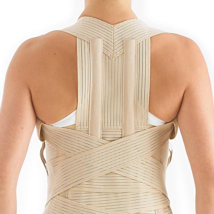
- Doctors recommend wearing special fixing corsets on the sacro-lumbar region. This helps prevent the development of complications, reduces the risk of injuries.
- An integral part of the treatment is special gymnastics. A set of exercises should be selected by an experienced physiotherapist. Regular physical activity helps to strengthen the muscle corset and relieve stress from the spine.
- Massage is also effective, which helps relieve spasm, strengthen muscles, improve blood circulation.
- Procedures such as electrophoresis with novocaine and paraffin baths help relieve soreness, warm up muscles, and eliminate cramping and discomfort.
- Acupuncture sessions will help to cope with unpleasant sensations.
Features of surgical intervention
Unfortunately, sacralization of the L5 vertebra is far from always amenable to conservative therapy. The decision to conduct surgery is made by the doctor if the following indications are available:
- severe pain that spreads to the back and limbs;
- compression of the nerve roots, which leads to a violation of normal urination and defecation;
- loss of sensation in the limbs, which is again associated with compression or nerve injuries.
There are several methods of surgical correction:
- excision of the apices of the transverse processes of the vertebrae;
- surgical separation of border vertebrae;
- placement between the vertebral bodies of a special transplant, which replaces the missing intervertebral disc and performs its functions.
In most cases, operations proceed without any complications. Naturally, a relatively long rehabilitation period follows, which includes some methods of conservative therapy.