The skin is considered the largest organ in humans. It protects tissues and organs from physical damage, high temperatures and chemicals. As you know, skin color is different. First of all, it depends on race, genetic characteristics of the organism, as well as on environmental conditions. The color of the skin is provided by special cells - melanocytes. Normally, they are located evenly in the basal layer of the epidermis. However, in some areas a large accumulation of melanocytes is found. This is visible to the naked eye. Areas of skin on which there is an accumulation of melanin cells are called pigmented nevi. Such formations are familiar to everyone. They are called moles or age spots. The size of such formations may vary. They also differ in color intensity: from light brown to deep black.
Pigment nevus: what is it, photo
It's no secret that almost every person has moles on the skin. Nevertheless, not everyone knows that “pigmented nevus” is a synonym for these formations. However, this concept is much broader. It includes not only small moles, but also age spots, reaching large sizes. Nevi can be located on the skin, as well as on the mucous membranes and even on the iris of the eye. Formations consisting of melanocytes differ in size, thickness, shape and color. So what is pigmented nevus? Photos of such formations can be found in the specialized literature on dermatology, cosmetology or oncology. Viewing images of various nevi will help to get an idea of their types. Despite this, in order to verify the origin of the mole, it is necessary to consult a specialist.
In most cases, pigmented skin nevi appear in early childhood. Small brown formations that slightly rise above the surface of the epidermis are called moles. They grow almost imperceptibly and do not bother the baby. Nevuses include birthmarks. These formations have a larger size and various shapes. They rarely rise above the surface of the skin. A baby is born already with these age spots, and they grow with it.
All nevi are composed of pigment - melanin, which gives color to our skin, iris and hair. The amount of this substance varies. The pigment content in the body is higher in dark-haired and dark-skinned people. Accumulation of melanin in one place leads to the formation of nevi. They can be located anywhere, including on the internal organs and muscles. Pigmentary nevus is a benign neoplasm that normally does not bother a person in any way. Most often, birthmarks cannot be treated if they do not cause aesthetic discomfort. A few years ago, it was believed that small pigmented formations on the face, on the contrary, give beauty. It is now known that not all moles are safe. In cases where there is a risk of malignancy of a benign neoplasm, it should be removed.
Causes of nevi
All pigmented nevi of the skin can be divided into congenital and acquired. This classification is not scientifically based, since it is still unknown when exactly the accumulations of melanocytes form. This division is based only on the appearance of nevi. If skin areas become dark in early childhood, then the formation is considered congenital. Acquired nevi appear in adolescents or adults.
The causes of congenital age spots are unknown. It is believed that pathological migration of melanoblasts occurs even during fetal development. Risk factors include illness during pregnancy, medication, or other chemical influences, as well as hormonal imbalances. A possible cause of the appearance of congenital nevus is a genetic predisposition. This explains the appearance of "Mongolian spots" in children of Asian origin.
Acquired nevi are considered more dangerous, since the frequency of transformation of these neoplasms into a cancerous tumor is higher. However, according to other sources, all pigmentation sites are formed at the stage of fetal development, and their appearance indicates an adverse effect of provoking factors. Regardless of this, the following causes of nevus occur:
- Hormonal changes in the body.
- Infections of the skin.
- Solar insolation.
- Visit to the solarium.
- Skin damage.
In fact, the selection of such provoking factors is quite justified. Hormonal imbalance develops during puberty and pregnancy. During these periods, the frequency of moles is much higher. In addition, age spots often appear in women using hormonal contraceptives.
Chronic skin pathologies (atopic dermatitis, acne) and physical damage often provoke the appearance of age-related pigmented nevuses on the face, neck and shoulder. The main reason for the appearance of moles is considered solar insolation. Exposure to ultraviolet radiation not only affects the occurrence of pigmentation, but also increases the risk of malignancy of formations. Therefore, people with problem skin should not be exposed to direct sunlight for a long time.
Classification of pigmented lesions
According to the histological structure, about 50 varieties of nevi are distinguished. Of these, the most significant are 10 types of benign formations that differ in origin and clinical picture. Doctors distinguish 2 main groups of nevuses, each of which includes several types of age spots. The classification is based on the risk of malignancy of education. The first group includes melanoparous nevus. The risk of malignancy of such formations is small. These include the following types of age spots:
- Papillomatous nevus. It differs in that it resembles melanoma in appearance: it has an uneven tuberous surface and rises above the surface of the skin. The hair on this formation continues to grow, but change color. Papillomatous nevus has a dark brown tint. The localization of such a mole is the trunk, limbs and scalp.
- Mongolian spot. It has a different shape and large size. This congenital pigmented nevus is found in most children of the Mongoloid race. It does not rise above the epidermis and disappears on its own by the age of 20.
- Halonevus - refers to acquired benign skin lesions. Has an oval or rounded shape. The peculiarity of this mole is that there is a light rim around it. A similar formation can be located on any part of the skin or on the mucous membranes. With age, the mole brightens and disappears.
- Intradermal pigmented nevus. It is characterized by small size and a peculiar localization: the neck and folds of the skin. Appears most often in the puberty. Under the influence of adverse factors, it can be malignant in melanoma. The complex pigmented nevus has a similar structure. It is a small pigmented formation resembling a papule.
- Fibroepithelial nevus. This formation consists of connective tissue and is rarely malignant. It protrudes significantly above the surface of the skin, has a light brown or pinkish tint. Such a mole has a rounded shape and a smooth surface. Education can be congenital, but more common in older people.
Melanoparous nevi are rarely malignant, but they require observation. The lack of growth of such moles indicates a favorable prognosis.
Melanopausal skin nevi
Melanopausal nevi include benign skin neoplasms, the likelihood of malignancy of which is high. Therefore, they require constant monitoring or radical treatment. The following formations belong to similar age spots:
- Blue Nevus of Yadassonne - Tiche. Despite the fact that it consists of differentiated pigment cells, pathology refers to precancerous conditions. Blue nevus has a small size (up to 1 cm) and slightly protrudes above the surface of the epidermis. In some cases, it is represented by a nodule located in the thickness of the skin. The formation has a purple or dark blue color.
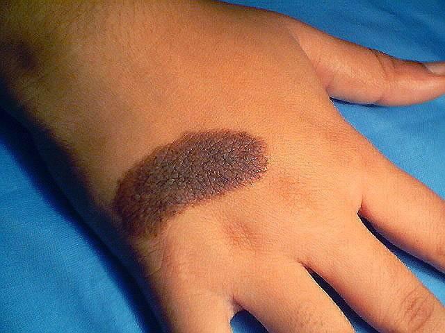
- Border pigmented nevus. Refers to congenital formations. A similar mole protrudes above the surface of the skin and has a dark color. The color of the pathological area may be purple, brown or dark gray. The size of the nevus does not exceed 1.2 cm. The name of this formation is due to the fact that the pigment cells of which it consists are located on the border of the epidermis and dermis.
- Giant nevus. This pigment spot is large (more than 20 cm) and can occupy a significant area of the body. The giant nevus has a rough surface and dark color. On the pathological area of the skin, enhanced hair growth is noted.
- Nevus Ott. A disease characterized by the presence of bluish spots on the skin of the face, lips, mucous membranes of the eye. It is often a birth defect, but can also occur in adolescence. The risk of developing such a disease is significantly increased among the representatives of the Mongoloid race.
- Nevus Clark. It is characterized by the asymmetry of the contours, a flat surface and various colors. The size of the skin defect ranges from 5 mm to 6 cm. The nevus can be located in the back, along the back of the thighs, or around the genitals. It refers to dysplastic formations, which are highly likely to transform into oncological pathology.
Melanopausal nevi is an extensive group of pathological conditions requiring diagnosis and treatment. In some cases, when the removal of education is impossible, constant prevention of malignancy is necessary.
Nevus eyes: features
Accumulation of melanocytes is observed not only on the skin, but also on the mucous membranes. An example is the pigmented nevus of the eye. Another name for this formation is a benign tumor of the choroid. It refers to congenital pathologies, however, it begins to appear only in 10-12 years. This is due to the fact that at this age there is an increased formation of pigment. There are 3 types of nevi of the choroid:
- Stationary.
- Progressive.
- Atypical.
All of them belong to benign tumors of the eye, however, under the influence of provoking factors, they tend to malignancy. Like skin lesions, choroid tumors are characterized by discoloration. So, what is it - pigmented nevus of the eye? Photos of such tumors are presented in abundance in the literature for ophthalmologists and oncologists, as well as on medical sites. Nevuses are small spots in the eye, the color of which differs from the color of the iris.
Types of Choroid Tumors
The stationary nevus of the eye is characterized by clear or feathery contours. It has a greenish or gray color. The shape, size and color of the formation does not change throughout life. Such tumors are practically not malignant.
Progressive nevus is characterized in that it has a yellow rim around the main accumulation of pigment. The color and shape of the defect may vary. In addition, such nevi increase in size, which increases the risk of vascular compression and a decrease in visual fields. Therefore, with this type of pathology, the supervision of a doctor is required.
Atypical nevi have an unfavorable prognosis. Therefore, with the slightest change or growth, urgent surgical treatment is required. Such formations are distinguished by light color and are accompanied by visual impairment.
Diagnosis of pigmented tumors
In case of changes in the nevus or its appearance, you should consult an oncologist. He will be able to make a differential diagnosis between various skin neoplasms and choose treatment tactics. An important study is dermatoscopy, which allows you to see the age spots under high magnification. If a tumor is suspected to be malignant, a full laboratory diagnosis, chest x-ray and abdominal ultrasound are performed. To establish the histological type of nevus, a wide excision of the formation is performed. A biopsy is carried out only in emergency situations, as it can lead to malignancy and the spread of tumor cells.
Pigment nevus: treatment
A photo depicting a nevus can be found in many sources of relevant subjects. Nevertheless, only a doctor can finally determine what kind of tumor it is. Treatment of pigmented nevus is not always carried out. In cases where the mole is harmless and does not cause cosmetic discomfort, dynamic observation is indicated. Nevi, which are often injured, are subject to removal. You can remove the formation using liquid nitrogen or a laser. If there is a risk of malignancy, surgical removal of pigmented nevi is required. At the same time, they recede from the spot by 2 cm and capture healthy tissue. The resulting material is sent to a histological laboratory.
Possible complications of nevus
The main complication of nevi is the transformation of normal pigment cells into melanoma. The following signs of malignancy are distinguished:
- A sudden increase in education.
- Bleeding or ulceration.
- Color change moles.
- Painful sensations.
- Itching and burning.
If you have any of these symptoms, you should immediately contact an oncologist and remove the formation. Only timely surgical treatment will help avoid cancer.
Prevention of malignancy of skin lesions
To prevent age spots from becoming malignant, all possible risk factors should be excluded. This especially applies to exposure to the sun and trauma to the nevus. People who have moles on their bodies are not recommended to sunbathe and visit the solarium.
The methods of prevention of melanoma include dynamic monitoring by an oncologist, as well as the timely removal of dangerous nevi.