The fact that microbes surround us was discovered by the Dutch scientist Levenguk. Pasteur later managed to establish a connection between them and many diseases. Microbes appeared on Earth one of the first and were able to perfectly survive to this day, populating almost every corner of the globe. They are found in hot vents of volcanoes and in permafrost, in waterless deserts and in the waters of the oceans. Moreover, they are perfectly settled in other living organisms and thrive there, sometimes bringing their master to death.
How were microbes discovered?
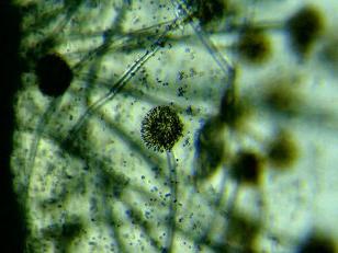
Anthony Levenguk invented a microscope and with his help he liked to examine what is impossible to see with the naked eye. It was the year 1676. Somehow, the inventor decided to find out why the tincture of pepper burns the tongue, looked at its solution through a microscope and was shocked. In a drop of matter, as if in some kind of fantasy world, hundreds of sticks, balls, spirals, hooks were spinning, sliding, pushing or lying motionless. This is what microbes look like under a microscope. Levenguk began to examine under the microscope everything that fell under the arm, and everywhere he discovered hundreds of previously unknown creatures, called animalocalcules by him. The scientist scraped a plaque from his teeth and also looked at him with the help of a device. As he later wrote, there were more animals in the plaque than there were inhabitants in the whole Kingdom. These simple studies laid the foundation for a whole science called microbiology (photo of a fungus on bread).
Microbes - who or what?
Microbes are a huge group of simple microorganisms that combines in their ranks non-nuclear creatures (bacteria, archaea), and having a nucleus (fungi). There are countless numbers of them on Earth. Bacteria alone, there are about a million species. According to a number of signs, they are classified as
living organisms. Many are interested in how microbes look under a microscope. Their appearance is quite diverse. The sizes of microbes range from 0.3 to 750 micrometers (1 micron is equal to a thousandth of a millimeter). In shape they are round like a ball (cocci), rod-shaped (bacilli and others), twisted into spirals (spirillos, vibrios), similar to cubes, stars and bagels. Many microbes have flagella and villi for more successful movement. Most of them are unicellular, but there are also multicellular ones, for example, fungi and the blue-green algae bacterium (photo of the mold bacteria).
Living conditions and habitat
Most of the microbes known today exist in environments with a moderately warm temperature. 40 degrees and above they can withstand no more than an hour, and when boiled, they die instantly. Also, radiation and direct sunlight are harmful to them. However, there are extremes among them that can withstand even + 400 degrees Celsius! And the bacterium flavobactin lives in the stratosphere, not afraid of either cold or cosmic radiation.
All bacteria breathe. Only one needs oxygen for this, and the other needs carbon dioxide, ammonia, hydrogen and other elements. The only thing all microbes need is a liquid. If there is no water, even mucus will do. These are microorganisms living in the body of animals and humans. It is estimated that in each of us there are approximately 2 kg of microbes. They are in the stomach, intestines, lungs, on the skin, in the mouth. Microbes under the nails are very numerous (this is clearly visible under the microscope). During the day, we take up a lot of objects with our hands, putting the microbes on them in our hands. Most microbes destroy ordinary soap, but under the nails, especially long ones, they linger and successfully multiply (photo of bacteria on the skin).
Food
Microbes, like people, feed on proteins, carbohydrates, mineral supplements, and fats. Many of them “love” vitamins.
If you look at the microbes under a microscope with a good increase, you can consider their structure. They have a nucleoid that stores DNA, ribosomes that synthesize proteins from amino acids, and a special membrane. Through it, microbes absorb food. There are autotrophic microbes that absorb the substances they need from inorganic compounds. There are heterotrophic ones that can only eat ready-made organic substances. This is all known yeast, mold, putrefactive bacteria. Human food for them is the most desirable medium. There are paratrophic microbes that exist only at the expense of the organics of other living creatures. These include all pathogenic bacteria. The bulk of microbes, with the exception of halophiles, cannot exist in a medium with a high concentration of salt. This feature is used for salting food (photo gonorrhea bacteria).
Breeding
Incredibly, some types of microbes have a sexual process, albeit in the most primitive form. It consists in the transfer of hereditary genes from parent cells to offspring. This happens through the contact of "parents", or the absorption of one by another. As a result, the “baby” microbes inherit the characteristics of both parents. But most germs and bacteria reproduce by dividing by transverse constriction or budding. Observing the microbes under a microscope, you can see how some of them have a small process (kidney) at one end. It rapidly increases, then it separates from the mother's body and begins an independent life. In this way, the “mother” microbe can produce up to 4 offspring, then it dies (photo Helicobacter pylori, causes gastrointestinal ulcers, cancer).
How are germs different from viruses?
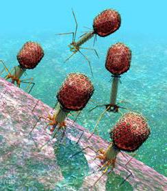
Some people think viruses and germs are the same thing. But this is wrong. Viruses, being the most numerous form of life, belong to organisms that live only at the expense of others. If we can see microbes under a microscope or even through a magnifying glass, then viruses that are a hundred times smaller than bacteria can only be examined with powerful electron microscopes. All viruses are parasites that cause diseases in humans, plants, animals, and even microbes. The latter are called bacteriophages. On Earth, there are many more than bacteria. For example, in a spoon of seawater there are about 250 million of them. Because sea water is useful, the bacteria contained in it kill bacteriophages. Attached to the body of the bacterium, they destroy its shell and penetrate inside. There, viruses begin to produce their own kind, as a result of which the host cell dies. Virusophages behave in the same way. This property is used in medicine in the production of antibiotics (pictured are bacteriophages).
Germs friends
Amazingly, only a tenth of our trillions of cells are actually human cells. The rest belong to bacteria and germs. This photo of microbes under a microscope represents bifidobacteria. They help us digest food, protect against pathogenic microbes, and produce amino acids. Our gastrointestinal bacteria are of great benefit. However, only as long as their number is strictly balanced. As soon as there are more bacteria than necessary, a person develops various diseases, from dysbiosis to stomach ulcers.
Sour-milk bacteria that “make” kefir, cheeses, yogurt for us are also useful. Bacteria are also used in the production of wine, yeast, environmental herbicides, fertilizers and much more.
Our worst enemies
In addition to the “good” microbes, there is a huge army of “bad” pathogens. These include plague stick, bacteria diphtheria, syphilis, tuberculosis, cancer, etc. There are trillions of bad microbes around us. They are everywhere, but there are especially a lot of them in public places - on handles in public transport, on money, in public toilets. Microbes on the hands under the microscope, if you look at them after returning from the store, are simply teeming. Therefore, hands need to be washed often, but without fanaticism. It is undesirable to use antibacterial agents, as this leads to dry skin and weakens the immune system.
A shocking spectacle is also caused by microbes on the teeth under a microscope. They get into our mouths with food, with kisses, with breathing. It is hard to say how many of them are in the oral cavity, if only up to 100 million parasites can be counted on a toothbrush. Especially if the toothbrush is stored in the same room with the toilet. Microbes in the mouth are the culprits of caries, periodontal disease, and infectious diseases. Their activity can be prevented by regular brushing of teeth and tongue, and after each meal - by rinsing the mouth with bactericidal drugs.