The disease, which will be discussed later in the article, is usually referred to the group of complex hereditary pathologies. Its main characteristic is the formation in the body of multiple tumors originating from tissues of various organs. What organ is most affected will depend on how Hippel-Lindau syndrome manifests itself.
However, there is a known case involving eight patients originating from the same family when the only symptom of pathology was polycystic kidney disease. The diagnosis in this case was established by identifying a genetic defect (mutation).
Hippel-Lindau disease is classified as a rare pathology, and its frequency of occurrence is one case per 36,000 newborns.
The mechanism of development of pathology
Hippel-Lindau disease is based on a generalized disorder of a hereditary nature, characterized by excessive growth of capillary tissue and the appearance of a large number of tumor neoplasms.
The disease is transmitted to the next generation in an autosomal dominant manner, that is, if one of the parents has a gene coding for the pathology, the probability of the offspring being sick is 50%.
A defect (mutation) leads to suppression of the gene, which is a suppressor of tumor growth.
Manifestations - oncological processes
The main manifestations of Hippel-Lindau syndrome are tumors of the pituitary gland, cerebellum, spinal cord, retina, and internal organs. We list the possible neoplasms:
- Hemangioblastomas - are localized in the cerebellum or retina; come from nerve tissue. CNS neoplasms have a frequency of occurrence in patients with Hippel-Lindau disease of at least 60%. The average age of patients in whom this type of tumor of the central nervous system was detected is 42 years.
- Angiomas are neoplasms of vascular origin that are localized in the spinal cord.
- Cysts These are neoplasms that have a cavity filled with fluid inside. Such elements can form in the liver, kidneys, testicles, and pancreas. In particular, cysts of the kidneys and pancreas are found in 33-54% of cases.
- Pheochromocytoma is a tumor originating from the cells of the adrenal medulla. The frequency of occurrence is about 7% of cases. The average age of patients with pheochromocytoma is about 25 years.
- Cell carcinoma. In particular, the average age of patients with this tumor is 43 years.
- In men with Hippel-Lindau disease, benign neoplasms in the testicular region (for example, papillary cystadenoma) may develop.
- In women, there is a tumor lesion of the uterine ligaments.
Criteria for diagnosis
A diagnosis of von Hippel-Lindau disease can be made if at least one of the symptoms presented in the following list is found. But this is possible if at least one of the family members has a history of hemangioblastoma of the central nervous system, hemangioblastoma of the retina, cystic formations of the kidneys or adenocarcinoma:
- hemangioblastoma, which is localized in the tissue of the cerebellum, as well as in other parts of the central nervous system;
- hemangioblastoma of the spinal cord;
- hemangioblastoma with localization in the bone marrow;
- retinal hemangioblastoma;
- cystic lesion of kidney tissue;
- renal adenocarcinoma;
- pancreatic cystic formations;
- cystadenocarcinoma;
- pheochromocytoma;
- epididymal adenoma.
It should be noted that medical specialties, whose representatives are involved in the problem of Hippel-Lindau syndrome, are neurology, ophthalmology, urology, and therapy.
CNS hemangioblastoma
The most commonly reported manifestations of von Hippel-Lindau syndrome are the development of hemangioblastoma of the cerebellum, spinal cord, or retina. A tumor developed on the cerebellum has the following manifestations:
- Headaches bursting in nature.
- Nausea, vomiting.
- Uncertainty when walking, due to impaired coordination.
- Dizziness.
- Impairment of consciousness (characteristic of the later stages of pathology).
With the localization of hemangioblastoma in the spinal cord, the main clinical symptoms are decreased sensitivity, paresis and paralysis, impaired urination and defecation. Pain is noted only occasionally.
This type of tumor is classified as slowly progressive. And the most informative for diagnostic and control examination methods are magnetic resonance imaging (MRI), enhanced by contrasting the study area.
The only effective way to treat cerebellar hemangioblastoma today is its surgical removal. The use of radiation and drug methods did not show a convincing positive result.
After microsurgical removal, hemangioblastoma, as a rule, does not recur, but other neoplasms may appear. In the surgical treatment of this tumor, it is necessary to take into account its multiplicity, which is characteristic of Hippel-Lindau disease.
Retinal angiomatosis
For ophthalmic localization of the pathological process, a characteristic feature is a triad of signs:
- the presence of angioma;
- fundus vasodilation;
- the presence of subretinal exudation (accumulation of fluid - an inflammatory product - under the cornea), which can cause the development of exudative retinal detachment at an advanced stage of the disease.
For the initial stages of the disease, a decrease in pigmentation on the fundus, an increased crimp of the blood vessels, are typical. With pressure on the eyeball, pulsation of both arteries and veins is noted.
At later stages, vasodilation and tortuosity of blood vessels progress, possibly the formation of aneurysms and angiomas, which have the characteristic appearance of rounded glomeruli, is a specific sign of Hippel-Lindau disease.
Pheochromocytoma
With adrenal gland damage, Hippel-Lindau disease is manifested by the formation of pheochromocytoma. This neoplasm, originating from the substance of the adrenal medulla, has, as a rule, a benign character. It is diagnosed in most cases at the age of about 30 years and is characterized by two-sidedness, multiplicity of nodes and the ability to switch to tissues located nearby.
Pheochromocytoma is manifested by the following symptoms:
- Hypertension resistant to antihypertensive therapy .
- Blood pressure related cranialgia.
- Pale skin.
- Tendency to hyperhidrosis.
- Tachycardia.
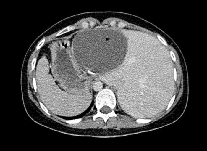
Pancreatic cancer
The oncological pathology of the pancreas is variable: tumors can be either benign or malignant. And in structure, these are either cystic formations or neuroendocrine tumors.
Various oncological processes affecting the pancreas are found in half of patients with Hippel-Lindau disease. Symptoms are usually associated with impaired pancreatic function.
The average age of patients at the time of detection of neoplasms is 33-35 years. In the case of a malignant process, the metastatic process is directed to the liver.
Endolymphatic sac tumor
Developing with the named syndrome, this oncological process always has a benign course and is manifested as follows:
- Hearing impairment, up to complete hearing loss.
- Dizziness.
- Discoordination disorders.
- Noise in ears.
- Paresis of the muscles of the face.
Diagnostics
Preliminary diagnostics are carried out on the basis of a general and ophthalmological examination of the patient, a medical history, including hereditary-family history.
Very informative is a blood test that reveals the content of glucose and catecholamines in it.
It is advisable to visualize the neoplasms using magnetic resonance imaging (MRI).
A source of valuable information for the early diagnosis of eye pathology is fluorescence angiography. This diagnostic technique helps to identify the initial manifestations of the pathology in the vasculature of the fundus, which are not recorded during ophthalmoscopy (such as telangiectasia, newly formed vessels). In the case of the progression of the pathological process, this technique allows you to identify the tumor-feeding vessel much earlier than ophthalmoscopy.
A complete comprehensive examination consists of the following diagnostic procedures:
- Computed and magnetic resonance imaging. Thanks to the use of these methods, the number of diagnosed diseases in the early stages of the disease has significantly increased. And in this case, they are often amenable to therapy.
- Ultrasound tomography.
- Angiography is a source of objective information about which organ the neoplasm comes from.
- Pneumoencephalography.
It is important to know that if a tumor process is detected in one of the organs, it is necessary to conduct a full examination to exclude the pathology of other areas.
Treatment
General approaches to the treatment of Hippel-Lindau syndrome are consistent with the principles of cancer therapy. Therapy should be comprehensive and combine surgical and radiation methods. Each of them is used strictly in accordance with the indications, contraindications, features of a particular tumor and the condition of the patient.
Medications are mainly used as symptomatic therapy to correct certain functions of individual organs and the general condition of the patient.
Forecast
With timely diagnosis and correctly selected treatment, the development of the disease, as well as its negative effect on the body, can be controlled. A prerequisite is the diagnosis and the beginning of treatment in the early stages.
However, in some cases, despite the efforts made, the prognosis remains unfavorable for the patient due to the high risk of complications.