X-rays of the lungs have recently become widely used in medicine. The reason for viewing the lungs in the picture can be very many symptoms. So, if lung cancer is suspected , an x-ray is simply necessary. Harmful tumors or other diseases (tuberculosis, foreign bodies, inflammation, fungal diseases) in this area can be the cause of many health problems. Among the long list of the latter, a number of fairly common symptoms can be distinguished: hemoptysis, dry cough, pain in the lungs, weight loss, general weakness, and many others.
Thus, a lung x-ray is simply an indispensable procedure for the diagnosis of diseases of the main respiratory organs. As you can understand, the production of lung images is based on the same principle as with conventional bone x-rays, that is, with the use of
x-rays penetrating into a certain area
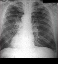
. It has only some of its differences. The fact is that in a healthy state, the lungs practically do not restrain the above-mentioned rays, in the picture you can see their "purity" even without special knowledge. If the lungs do not have pathologies, then their image can be observed on the image in the form of two fields on which the vessels are traced. In the case of tumors or other diseases, the rays are delayed, which, of course, is displayed in the picture. One way or another, but to consider the disease without deep medical knowledge is usually difficult, the help of a doctor is necessary in any case.
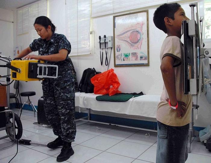
An X-ray of the lungs does not require preliminary preparation of the body and does not foresee any contraindications. The procedure itself takes place, as a rule, in two stages. First, the patient leans against the device with his hands spread apart on the sides, and a picture cassette is placed on the opposite side. Next, the same procedure is carried out, only the diagnosed is placed sideways. The result is two shots. In rare cases, X-ray of the lungs is applied in a half-turn (45 degrees). An X-ray of the lungs is also possible, in which the picture is not taken, and with the help of the appart, the region of the lungs is viewed in X-rays.
People are often interested in the difference between lung x-rays and fluorography. And if it is generally known where to do fluorography, then pulmonary x-ray is often not possible to do in an ordinary state hospital. Here you have to use the services of private offices. Despite similar tasks, lung X-ray and fluorography have many differences. First, the differences relate to the research methodology. If a fluorographic image before a preliminary examination involves a rather complex processing system, then a lung x-ray is carried out simply by high-quality photographing of the lungs and direct examination. Secondly, the size of the obtained images is significantly different, fluorography involves obtaining many times smaller images. Thirdly, another drawback of fluorography is its poor informational content, while the lung X-ray provides for a more detailed picture. Fourth, fluorography can, in principle, be considered as a preventive, technically simple diagnostic method.
We can conclude that X-ray is a very important element in the diagnosis of various
lung diseases and the fight against them, which is much superior to the widely used and outdated lung fluorography.