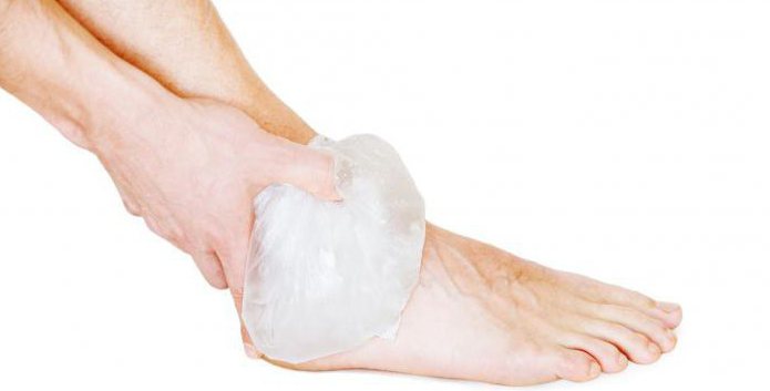
Not a single person is insured against injuries and injuries. In addition, there are separate parts of the body that are at greatest risk of developing various pathologies. One of these is the ankle joint. Dislocation, the appearance of swelling, soreness and other unpleasant symptoms serve as an alarm signal, indicating the need for seeking medical help in the near future.
Before starting treatment, specialists will prescribe the patient to undergo diagnostic procedures. A timely examination will help identify the cause of the pathological condition of the ankle joint. MRI is an ultramodern and most informative method of research, giving a detailed picture of internal structures and assessing the state of muscles and soft tissues. It is quite difficult to establish an accurate diagnosis without tomography. In addition, the causes of the development of pathology can be a variety of factors.
Advantages of magnetic resonance imaging in joint pathology
MRI scan of the ankle has a considerable advantage over other types of research. Its advantages lie in the possibility of obtaining high-quality images, which allows specialists to detail the image with a certain accuracy. In the conclusions of the diagnostician, you can find an objective assessment of the condition of the bones, muscle system, soft tissues, ligaments. During the procedure, specialists have the opportunity to visually examine the characteristics of blood flow in a particular clinical case.
With the most complex injuries and injuries of the leg, magnetic resonance imaging is the only type of study that guarantees an accurate result. In addition, an MRI of the ankle joint does not take much time: scanning lasts about 20-30 minutes, when using contrast - no more than an hour. The procedure for the patient takes place without any side effects and is absolutely painless, therefore, upon its completion, the researcher can return to his usual way of life. MRI is a non-invasive method that allows you to look inside the human body and get maximum information about its condition.
Following the provision of first aid for injuries of the ankle joint, the victim is sent for diagnosis. The study is prescribed by a surgeon or traumatologist. Without preliminary MRI, it is impossible to carry out surgical treatment, and therefore tomography is mandatory for patients who are shown surgery.
Indications for ankle MRI
Other indications for undergoing the research procedure are the following conditions and diseases:
- the presence of neoplasms of unknown histology (if a benign or malignant tumor is suspected);
- fractures and dislocations of the ankle joint;
- sprains, tears of ligaments and tendons;
- pain syndrome of unknown etiology in the lower limb;
- swelling and limited motor activity of the foot;
- pinched nerve in the foot.
In addition, some chronic conditions are the most common reason for MRI scans. For example, tomography is recommended for arthrosis or arthritis of the ankle joint, the symptoms and treatment of which require periodic monitoring by qualified doctors. The study reveals problems even when other research procedures have shown doubtful results, including those related to circulatory disorders in the limb.
Thus, magnetic resonance imaging is a comprehensive diagnostic method, notice the development of the pathological process in its early stages and begin to eliminate deviations as early as possible.
Contraindications and Precautions
Despite the safety of the procedure, in some cases it is recommended to refrain from MRI. The body examination technique, which implies the introduction of a contrast medium, as a rule, has more contraindications than a standard scan. Especially careful when undergoing a diagnostic procedure should be suitable for patients who have a history of a tendency to allergic reactions. During pregnancy, as well as with breastfeeding, the administration of a contrast medium is unacceptable even in minimal doses. Gadolinium, which is part of it, adversely affects the work of the kidneys, so it is undesirable to resort to this diagnosis to people suffering from renal failure.
In connection with the features of an MRI of the ankle joint and the principle of operation of the equipment, it is important to adhere to other restrictions:
- Patient weight should not exceed 120 kg.
- It is better to conduct a study for people suffering from claustrophobia or mental disorders in an open type tomograph.
- Do not conduct a study for patients with metal structures in the body. Whether it's a heart valve, a pacemaker, an insulin pump, a prosthesis or just a piercing - any iron elements can distort the diagnostic results.
- When the couch is immersed in the chamber, the patient's body is tightly fixed with retaining straps in order to avoid involuntary movements.
Can an MRI be done in childhood?
It is extremely rare to apply this research method to children, for which there are a number of reasons. In addition to the harm from the contrast medium, there are other points that impede the procedure at such an early age. Compliance with motor immobility, which is required with magnetic resonance imaging, is often an impossible task for children.
In case of acute need for diagnosis, the child is examined under general anesthesia. That is why it is more advisable to have an MRI only in cases where the expected benefits of the diagnosis exceed the potential risk to the baby’s health.
Informativeness of the procedure: what will the MRI show?
If the ankle joint is swollen, but the cause of the pathology remains unknown, MRI is prescribed in 90 percent of cases. Due to the versatility of the study, doctors receive detailed information about the condition:
- bones, since tomography allows you to visually examine the lower parts of the tibia, partially calcaneus and talus;
- ligamentous apparatus - tendons and ligaments, amenable to tears and stretching even without heavy physical exertion, often cause many years of pain;
- muscles - to confirm or exclude the inflammatory or infectious process of the soft tissues of the lower leg and foot;
- cartilage tissue - with the timely detection of areas of increased wear, cracks and other defects, the patient has every chance of preventing arthritis of the ankle joint.
Symptoms and treatment of most pathologies detected during the diagnostic procedure require a serious approach and further monitoring by specialists. Use this method of examining the limb and with suspicion of cystic or oncological formations.
In addition to the fact that MRI of the ankle joint indicates the presence of diseases, the procedure can be prescribed to the patient in order to monitor the effectiveness of the current treatment, as well as upon completion.
How to prepare for joint diagnosis?
If the patient was referred for the first time for research, then he certainly does not need to worry. Excited about the upcoming procedure, many vied with each other are looking for answers on how to do an MRI of the ankle joint. In fact, for the examination of this part of the body does not require any special training. The patient can go on an ankle scan by appointment immediately after receiving a referral from the attending physician.If it is necessary to administer gadolinium intravenously for MRI with contrast, it is advisable to arrive at the session a little earlier, since additional time will be required to distribute the substance throughout the body and on an empty stomach.
What is an ankle examination?
The procedure itself usually does not cause any difficulties. Briefly, the course of the study can be described as follows:
- Before entering the office, the patient removes all metal objects, including jewelry, jewelry, disconnects the mobile phone.
- In the early stages of pregnancy, an ankle scan without the introduction of a contrast agent is allowed, but before the procedure, the doctor must be warned about the special situation of the subject.
- Next, the patient will be asked to lie on the couch of the tomograph, the specialist will tightly fix his arms, legs, head with straps and rollers. To reduce the audibility (the device is working quite loudly), the patient may be given headphones.
- After this, the table is pushed into the chamber at such a distance that the foot and lower leg of the patient fall directly under the torsion path of the tomograph ring.
Research results and their interpretation: what to do next?
The procedure does not affect the sensation of the subject, even if his ankle joint is swollen. A doctor will be watching the progress of the study in the next office, who can be contacted at any moment and stop the scanning process in unforeseen circumstances.
Upon completion of the examination, the doctor will give the patient the results of the procedure. It is not necessary to independently search for the causes of pathologies in them and assess the severity of the ankle joint damage: the attending physician will decipher the study parameters.
What are the main diseases detected by tomography?
Focusing on the conclusion of the diagnostician, and based on the symptoms described by the patient, the specialist will be able to make an accurate diagnosis. With a fracture, soft tissue injury or partial rupture of the ligaments of the ankle joint, the patient will receive a comprehensive treatment, consisting, as a rule, of a course of restorative medicines and physiotherapy exercises. If a neoplasm is found, to determine its exact nature, the patient is sent for a biopsy to undergo a histological examination of the taken tissue samples.
The most common pathologies that can be used to detect MRI of the ankle joint are the following:
- arthrosis and rheumatic ailments (including infectious diseases, gout, juvenile rheumatoid arthritis);
- joint capsule damage, ligament rupture and sprain;
- joint hemorrhage;
- rupture of syndesmosis (more common due to dislocation of the ankle between the small and large tibia);
- abnormalities of embryonic development;
- chondrosarcoma - joint bone cancer;
- synovioma - a malignant tumor of the synovial membrane.
Which is better: MRI or CT? What is the difference between the two?
When choosing a diagnostic method, in most cases, doctors prefer an MRI. In some cases, computed tomography may be an alternative . It is difficult to outright determine which of the varieties of ankle joint scan is better and more effective, since the purpose of both procedures depends largely on the indications.
So, CT is carried out according to the principle of X-ray examination, i.e. transillumination of a part of the body by rays. Accordingly, this type of diagnosis is more effective for pathological changes in bone tissue. In the computed tomography image, the bones will be displayed using white shadows, and the remaining cavities will be indicated in dark color. This marking is effective to identify the location of the fracture, cyst, foreign body.
To see what condition the muscles of the ankle joint are in, it is preferable to stay on an MRI. Scanning of the joint itself, the ligamentous apparatus and the surrounding soft tissues will be more revealing than computed tomography.
How often can joints be scanned with MRI?
Despite stereotypical thinking and ideas about the dangers of magnetic resonance imaging for human health , this type of diagnosis is absolutely safe for the body. In the absence of contraindications and the observance of all necessary precautions, you should not worry: the study is not able to negatively affect the course of the disease, worsen the general condition of the patient. Unlike radiography and CT, MRI can be done much more often. Meanwhile, the decision on the need for diagnosis and the frequency of the procedure is made solely by a specialist.
How much does an ankle MRI cost in Russia?
Another point worth noting is the cost of ankle MRI. It is not easy to say unequivocally how much diagnostics cost in Russia, since the prices for carrying out the procedure can have significant differences in different regions of the Russian Federation. On average, the cost of tomography ranges from 4000-5000 rubles. If there is a compulsory medical insurance policy, patients still have the opportunity to undergo the procedure for free at the place of residence. Details should be checked with your doctor or insurance company that issued the policy.