T-cell lymphomas are most often diagnosed in elderly patients, but adolescents and children are also susceptible to this disease. Moreover, men suffer from it much more often than women.
What is T cell lymphoma?
Lymphoma is a malignant lesion of lymphoid tissue and other organs. In the case of T-cell lymphoma, the disease affects the skin and lymph nodes (that is, it is epidermotropic in nature). The main elements that are involved in the process of the onset of this neoplasm are T-lymphocytes (cells that normally provide an adequate immune response). T-cell lymphoma (photo below) can form as a result of the accumulation of pathological cellular elements in the tissues.
Causes and predisposing factors
- Heredity
- Immunodeficiencies.
- Long-term exposure to adverse factors: ultraviolet radiation, radiation, aggressive chemical agents.
- Age aspect (elderly patients).
- The presence of viruses in the body (HIV, Epstein-Barr, hepatitis C, B, T-lymphocytic virus), intestinal diseases.
- The presence of mutations in the immune system, organ and tissue transplantation, the appointment of immunosuppressive therapy.
All of the above factors can be aggravated (respectively, provoke the development of the disease) stresses, malnutrition, nervous overload, non-compliance with rest and work.
Classification
- T cell lymphoma of the skin. Such lymphomas, as a rule, develop as a result of various mutations of T-lymphocytes, leading to uncontrolled reproduction of the latter and, as a consequence, their spread in the epidermal layer. This type of lymphoma is characterized by the appearance of a polymorphic rash (plaque, tumor, spot, blister). Moreover, at first, patients complain of the appearance of scaly or itchy spots, which eventually transform into plaques, and subsequently into foci of the tumor.
- Peripheral T-cell lymphoma. This concept includes all lymphomas that have a T-cell or NK-cell origin (the exception is neoplasms from immature T-cells and T-lymphoblastic leukemia). Quite often, such lymphomas occur with damage to the blood, skin, bone marrow and internal organs, and diffuse infiltrative changes are observed in the lymph nodes.
- Angioimmunoblastic T-cell lymphoma. It manifests itself in the form of compaction of lymphoid tissue (during histological examination of the lymph nodes) by immunoblasts and plasma cells, with further complete transformation of its structure and the formation of pathological blood vessels.
- T-cell lymphoblastic lymphoma. A neoplasm based on pathological changes in T cells. T-lymphocytes do not mature, their nucleus acquires an irregular shape, they quickly and uncontrollably divide. This type of lymphoma is quite rare. When a similar pattern occurs, differential diagnosis with acute leukemia is performed.
- Non-Hodgkin T-cell lymphoma. It is a large group of neoplasms that consist of mature T-lymphocytes and do not belong to lymphogranulomatosis (i.e., Hodgkin's disease). Belonging to this group is determined by histological examination (with lymphogranulomatosis, specific cellular elements of Berezovsky-Shtenberg are found in the preparation, with non-Hodgkin lymphomas they are not).
Developmental stages
- The defeat of one lymph node.
- The spread of the tumor process into two lymph nodes that are located on one side of the diaphragm.
- Multiple lesions of the lymphatic structures on both sides of the diaphragm.
- Damage to the lymph nodes and internal organs, the presence of metastases in the kidneys, bone marrow, liver or organs of the gastrointestinal tract.
General symptoms
The presence or absence of certain clinical manifestations depends on the type of tumor and its location. However, there are symptoms that are inherent in almost all T-cell lymphomas:
- Sweating at night.
- Fast causeless weight loss.
- Mushroom mycosis.
- Low-grade indicators of body temperature.
- Frequent weakness and apathy.
Fungal mycosis
Mushroom mycosis is one of the most frequent manifestations that characterizes T-cell lymphomas. It occurs in seventy percent of patients with this disease and has three forms: classical, decapitated and erythroderma.
Classical fungal mycosis goes through three stages of development:
- Erythematous - according to clinical signs, it is similar to psoriasis, eczema and so on. Patients note the presence of oval or round spots, with clear boundaries, of various sizes, with a pink-purple color and peeling on the surface. Such elements are more often located on the face and body, gradually increasing in number and moving to other areas. Such rashes are not treatable and may not go away for a long time or suddenly disappear. In addition, spots can be combined with itching, which is not stopped by conventional means.
- Plaque-infiltrative - occurs a few years after the onset of the disease. In place of the above spots, dense plaques with round or irregular, but clear boundaries, purple in color with peeling on the surface. Brown spots or atrophy of the skin remain in place of the disappeared plaques . Itching intensifies, becomes unbearable. Rapid weight loss and fever are observed, lymphadenopathy may be present.
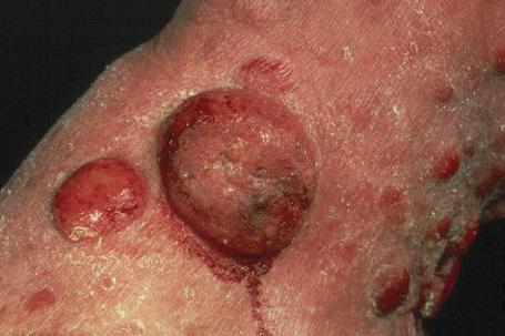
- Tumor - in place of plaques or on unchanged skin, the appearance of dense red-yellow painless tumor-like formations of a spherical or slightly flattened shape resembling a mushroom hat. The diameter of such formations ranges from one to twenty millimeters, and the number can be different. When a tumor collapses, painful ulcers form in its place, the depth of which can reach fascia or bone. Most often, the spleen, lymph nodes, lungs and liver are affected. Intoxication and weakness are growing. Patients from heart failure, amyloidosis, pneumonia die.
The erythrodermic form is characterized by the occurrence of growing painful itching, hyperemia and swelling, the appearance of merging erythematous-squamous spots, hyperkeratosis on the soles and palms, as well as hair loss. All lymph nodes in the axillary, inguinal, cubital and femoral areas are enlarged. When they are palpated, dense, elastic, painless packets of lymph nodes are found that are not fused with the surrounding tissues (skin). There is fever (up to 39 degrees), weight loss, night sweats and weakness. A fatal outcome is possible with exacerbation of intoxication and the addition of heart failure.
In the presence of a decapitated form, multiple plaques appear on healthy skin, eventually becoming malignant. The death of patients occurs within a year.
Clinical manifestations of cutaneous lymphomas
T-cell lymphoma of the skin often occurs against the background of long-existing and improperly treated dermatological diseases. Herpes viruses, radiation, sunburns and so on can provoke its occurrence. Similar lymphomas occur in two stages: primary (the process is localized in the dermis), secondary (when skin lesions are the result of penetration of lymphocytes from internal organs).
The main symptoms are:
- Polymorphic skin rash.
- Increased skin itching.
- The presence of small flat elements with a nodular shape.
- The appearance of yellowish plaques, subsequently transforming into foci of hyperpigmentation and atrophy (with plaque form).
- The clinical picture resembling psoriasis (with a small-type type).
- Dryness, swelling, hyperemia and peeling of skin areas (with erythrodermic type of disease).
Symptoms of peripheral lymphomas
The clinic of the disease depends on the stage and type of the pathological process.
An increase in lymphoid tissue in the cervical, inguinal and axillary regions is characteristic.
Patients complain of excessive sweating, severe weight loss, lack of appetite, general weakness, fever, and in the presence of an enlarged spleen and liver (which is common enough) - shortness of breath, periodic coughing and a feeling of heaviness in the stomach.
Diagnostics
- Inspection, collection of complaints and medical history from an oncologist.
- General analysis of blood and urine.
- Blood biochemistry, as well as analysis for antibodies to hepatitis viruses.
- A biopsy of the damaged lymph node (which allows verification of the diagnosis of t cell lymphomas).
- Ultrasound
- MRI
- CT
- Oncoscreening of the body.
- Immunophenotyped study.
- Molecular genetic analyzes.
- Cytogenetic analysis.
Therapy
The treatment of T-cell lymphoma depends on the form and stage of the process.
So, for example, for the erythematous stage of fungal mycosis, special antitumor therapy is not prescribed, but external corticosteroids and interferons are used.
Antitumor treatment boils down to chemotherapy and radiation therapy.
In accordance with the form of the disease (indolent, aggressive and highly aggressive), the duration and composition of the treatment, as well as rehabilitation, are determined.
Indolent lymphomas are not treated, but only regularly observed, if the process progresses, the tactics are changed.
In the treatment of aggressive forms, a combination of chemotherapeutic drugs and Rituximab (monoclonal antibodies) are used.
When it comes to highly aggressive lymphomas, drugs are prescribed that are used in the treatment of acute leukemia.
Chemotherapy is often combined with stem cell transplantation.
However, the most effective treatment for lymphoma is a thirty-day course of radiation therapy (according to the method of Elect Sinergi), and after taking chemotherapeutic agents.
Complications of chemotherapeutic agents may be: lung cancer, breast cancer, and so on. Radiation therapy may be complicated by the development of coronary atherosclerosis.
After successful treatment, compulsory rehabilitation is carried out.
T-cell lymphoma: prognosis
- With the timely detection of the disease (initial stage) and the appointment of adequate treatment, the five-year survival rate is eighty-five percent, and for pediatric patients - up to ninety percent.
- Regarding angioimmunoblastic lymphoma, the prognosis is disappointing. The average life expectancy in patients with a similar diagnosis is only two and a half to three years. Five-year survival is observed in only a third of patients.
- In the presence of a T-lymphoblastic form, the prognosis depends on the presence / absence of bone marrow damage. If the bone marrow is not affected, the outcome is usually favorable. Otherwise, improvement can be expected in only twenty percent of patients.
- The prognosis and survival of fungal mycosis depends on the form and stage of the process. So, with the classic form, survival is up to ten years, and the prognosis is poor in the case of pneumonia, and so on; the erythrodermic form is disarmed by the addition of various complications, as a result of which death occurs in two to five years; with a decapitated form, the prognosis is unfavorable, death occurs within a year.