As you know, any tissue of the body can undergo a malignant transformation. The hematopoietic system is no exception . Diseases of this tissue are divided into 2 groups: myelo- and lymphoproliferative neoplastic processes. The tumor pathology of the hematopoietic tissue is called hemoblastosis. This is the common name for all varieties of neoplastic processes. In most cases, hemoblastoses develop in childhood. However, some types of blood cancer are found exclusively in adults. Hematologist is involved in blood pathologies. He recognizes the type of hemoblastosis and prescribes the appropriate treatment. The main method for normalizing blood composition is chemotherapy.
What is hemoblastosis?
Like all oncological pathologies, hemoblastoses are characterized by the emergence and reproduction of immature cells. These can be undifferentiated elements of the hematopoietic or immune system. In the first case, the process is myeloproliferative in nature and is called leukemia. Reproduction of immature immune cells, some authors attribute to lymphomas, others - to hematosarcomas. Previously, similar blood cancers were called leukemia.
Unfortunately, hemoblastosis is one of the main causes of mortality from cancer. In the oncological structure of blood pathology, they occupy 5-6th place. Such tumors are especially common in preschool children. The main criteria of the disease include: intoxication, hemorrhagic, hyperplastic and anemic syndrome. Only after a qualitative blood test can a diagnosis of hemoblastosis be made. The ICD-10 code is assigned to each of the varieties of leukemia.
Causes of the development of diseases of the hematopoietic system
Blood cancer, like other neoplasms, usually develops suddenly, without any previous signs. Therefore, it is possible to recognize the cause of cell transformation in rare cases. Nevertheless, it is proved that the development of leukemia can be associated with provocative factors that preceded leukemia long before its onset. Similar reasons include radiation. Blood disease (hemoblastosis) often occurs after radiation exposure to the body. Therefore, etiological factors include ionizing and ultraviolet radiation, including frequent diagnostic procedures and therapy for other tumors. Among other causes of hemoblastosis, there are:
- Viral exposure.
- Congenital genetic abnormalities.
- Disorders of amino acid metabolism.
- Exposure to chemical carcinogens.
Epstein-Barr virus is found in some patients suffering from malignant lymphomas and hemoblastoses. This pathogen not only weakens the immune defense, but also activates the oncogenes present in the body. The role of retroviruses in cell degeneration is also being studied. Among genetic diseases, risk factors include: Klinefelter syndrome, Down, Louis-Bar. Chromosomal abnormalities and congenital metabolic disorders lead to impaired differentiation of myeloid and lymphoid cells.
Chemical carcinogens include some antibacterial and cytostatic drugs. The following medicines are an example: Chloramphenicol, Levomycetin, Azathioprine, Cyclophosphamide, etc. Therefore, the risk of leukemia is increased in people receiving chemotherapy for malignant tumors. Carcinogens are also found in enterprises using benzene and other harmful substances.
The mechanism of development of leukemia
The pathogenesis of all oncological diseases is based on a violation of the differentiation of cellular elements. Hemoblastosis is a pathology in which immature myelo- and lymphocytes appear in the blood. Disturbance of differentiation can occur at any stage of development of the progenitor cell. The earlier the violation occurs, the more malignant the disease. It is believed that under the influence of etiological factors mutations occur in genes. This leads to a change in the quality of chromosomes and their rearrangement.
All hemoblastoses (leukemia) are of monoclonal origin. This means that all pathological cells in the blood are the same in structure. Normally, the differentiation of blood cells goes through several stages. A precursor to all tissue elements is a stem cell. Ripening, it gives the rudiments for myelogenous and lymphoblasts. The former are converted to red blood cells and platelets. The second group of cells gives rise to elements of the immune system of the blood, that is, white blood cells.
Violation of stem cell differentiation leads to the fact that the composition of the blood changes completely. In the study, it is impossible to determine a single normal element. All of them are the same, therefore they cannot perform the necessary functions. This explains the fact that undifferentiated hemoblastosis is considered the most malignant cancer and has a worse prognosis. If maturation is impaired in the later stages, the cells partially or fully function. Therefore, the prognosis for highly differentiated cancer is more favorable. However, even fully matured cells differ in pathological division and displace other normal blood elements.
Varieties of hemoblastosis in adults and children
Given the pathogenesis of hemoblastosis, the disease is primarily classified according to the degree of differentiation of pathological cellular elements. Not only the clinical picture of the disease depends on this, but also the selection of the right treatment. Depending on what type of cells has undergone changes, myelogenous and lymphoproliferative hemoblastosis is secreted. Each of these groups is divided into acute and chronic leukemia. The first is considered more unfavorable due to the low degree of differentiation. To detect acute leukemia, it is necessary to confirm the presence of blast cells. With the myeloid type, the pathological substrates can be the precursors of monocytes, megakaryocytes and red blood cells. Acute lymphoid hemoblastosis is a serious illness that occurs in childhood. In this pathology, immune cells possess pathological activity. Among them are the precursors of B- and T-lymphocytes, as well as CD-10 and CD-34 antigens.

Chronic hemoblastoses are also divided into myeloid and lymphoid. The former are characterized by an increase in the number of neutrophils, basophils, eosinophils or their mature precursors. The number of blast cells in chronic myelogenous leukemia is small. In most cases, the disease develops against the background of genetic mutations. Chronic lymphocytic leukemia is more often diagnosed among the elderly male population. Sometimes pathology is inherited. A similar ailment is divided into the following groups:
- T cell leukemia.
- Paraproteinemic hemoblastoses.
- B-cell leukemia.
All of these pathologies relate to malignant immunoproliferative processes. Paraproteinemic hemoblastoses, in turn, are classified into the following:
- Heavy chain disease.
- Primary Valdenstrom macroglobulinemia.
- Myeloma.
A feature of these types of hemoblastoses is that they synthesize fragments of immunoglobulins (paraproteins). The most common form of this group of leukemia is myeloma.
The clinical picture in chronic blood neoplasms
How is hemoblastosis manifested? Symptoms of lymphoproliferative blood diseases are associated with impaired immunity. Patients with chronic leukemia complain of infections that occur despite treatment. Symptoms of lymphoid hemoblastosis also include severe allergic reactions that have not been previously observed. This is due to the restructuring of the immune system and its excessive activation. The clinical picture of chronic myelogenous leukemia depends on the stage of the disease. At the initial stage, the disease resembles an inflammatory process and is accompanied by a low temperature, poor health, and weakness. In the terminal stage, the following symptoms are joined: bone pain, lymphadenopathy, enlarged spleen and liver. With progression, patients are severely depleted, weight loss occurs, infections join.

Due to the predominance of certain types of cells in the blood, the growth of other elements is inhibited. As a result, anemia and thrombocytopenia may occur. A decrease in hemoglobin level affects the general condition of the patient. The patient becomes lethargic, the skin becomes pale in color, there is a decrease in blood pressure, fainting is noted. With thrombocytopenia, hemorrhagic syndrome develops. Its manifestations include various bleeding.
Symptoms of acute leukemia
Compared with the chronic form of the disease, acute hemoblastosis is more pronounced. Symptoms of this ailment quickly increase, and a person’s condition worsens noticeably. The following syndromes prevail in the clinical picture:
- Anemic.
- Hemorrhagic.
- Lymphoproliferative.
- Hepatosplenomegaly syndrome.
- Intoxication.
- Immune system syndrome.
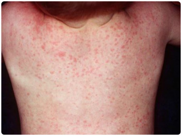
Due to inhibition of blood formation in patients, severe anemia is noted. This is especially pronounced with lymphoid leukemia. Despite the ongoing therapy, hemoglobin in patients remains low. The characteristic signs of anemia include pallor, severe weakness, dry skin, damage to the mucous membranes and a perversion of taste. Hemorrhagic syndrome is characterized by the appearance of red dots and spots on the skin (petechiae, ecchymosis). With a pronounced platelet deficiency, external and internal bleeding occurs, which leads to the progression of anemia.
Intoxication in patients suffering from hemoblastoses is manifested by a decrease in appetite, pain in muscles and bones, and constant weakness. Like any oncological process, blood cancer is accompanied by weight loss. Acute hemoblastosis is almost always accompanied by lymphadenopathy. Respiratory failure may develop from an increase in the size of the thymus. In addition to hypertrophy of all groups of lymph nodes, hepato- and splenomegaly are noted. The clinical picture of hemoblastosis in children is the same as in adult patients.
The progression of blood cancer leads to the defeat of almost all organs and systems. First of all, the testicles and kidneys suffer. The main complication of the disease is DIC, that is, a blood clotting disorder. Also, patients often suffer from joining infections that develop against the background of immunodeficiency.
Diagnostic methods for hemoblastosis
Acute hemoblastoses have the following diagnostic criteria: a decrease in hemoglobin at a normal color score, neutropenia, thrombocytopenia, and lymphocytosis in the KLA. The number of leukocytes differs depending on the type of disease. With lymphoid type hemoblastoses, their level rises sharply (tens or even hundreds of times). A decrease in the number of leukocytes can be observed in myeloproliferative blood cancer. The main diagnostic criterion for an acute pathological process is the presence of blast cells and the absence of intermediate elements. A similar blood picture is called leukemic failure. To confirm the diagnosis, a bone marrow analysis and a study are carried out for myeloperoxidase, chloroacetate esterase, SIC reaction.

Additional diagnostic criteria include: chest x-ray, cytogenetic analysis, ultrasound of the soft tissues and internal organs. The research algorithm for suspected chronic hemoblastosis is the same. In the KLA, a shift of the leukoformula to the intermediate elements of the blood (promyelocytes) is observed. Blast cells may be present in small numbers. In chronic myelogenous leukemia, the Philadelphia chromosome appears in the bone marrow. Serological examination and ELISA help confirm lymphoid type blood cancer.
Hemoblastoses: differential diagnosis of diseases
Based on only clinical data, it is difficult to make a diagnosis: hemoblastosis. After all, the manifestations of this disease are similar to other systemic pathological processes. Depending on the predominance of a particular syndrome, leukemia is differentiated with lymphogranulomatosis, aplastic and hemolytic anemia, and HIV infection. If respiratory failure comes first, the disease resembles a tumor of the mediastinum or lung. Only after a blood and bone marrow examination can hemoblastosis be distinguished from the listed diseases.
Treatment of acute and chronic leukemia
How is hemoblastosis diagnosed? The ICD-10 code is different for each type of leukemia. Acute myeloid blood neoplasm is assigned the code C92.0, and the chronic process is assigned the code C92.1. Lymphoproliferative leukemia is encoded as C91.0-C91.9. Depending on the diagnosis, a treatment regimen is selected. The main method is chemotherapy. For treatment, Vincristine, Endoxan, Doxilide, and Cytarabine are used. The choice of drugs depends on the type of hemoblastosis. Some regimens include the hormone drug Prednisolone. The treatment is aimed at the induction and consolidation (consolidation) of remission. Then prescribe drugs for maintenance therapy. Among them are the medicines Merkaptopurin and Methotrexate.
In addition to chemotherapy, radiation treatments and bone marrow transplantation are used. In some cases, splenectomy is performed.
Hemoblastoses: prevention and prognosis
It is impossible to predict the development of leukemia in advance, therefore, special methods of prevention do not exist. People with a history of cancer should take care of themselves from various radiation and chemical influences.
It should be remembered that some types of leukemia tend to be hereditary. Therefore, in the presence of blood cancer in relatives, it is necessary not only to lead a healthy lifestyle, but also to periodically take the KLA. An example is paraproteinemic hemoblastosis. The prognosis of the disease depends on the degree of differentiation of tumor cells and timely treatment. Five-year survival is 30 to 70 percent when remission and bone marrow transplantation are achieved.