Fibrous dysplasia is a fairly rare disease, which is characterized by systemic damage to the skeleton. At the same time, normal bone tissue is replaced by tumor-like connective cells.
The main reason for the onset of pathology is considered a gene mutation. However, doctors also call many other prerequisites for the development of anomalies, which may vary depending on the degree of its course.
Description
Fibrous dysplasia is a specific tumor-like formation of bone tissue. An anomaly develops even in childhood, it is characterized by a rapid progressive course. In the category of all bone tumors, this pathology is approximately 2%. In a fifth of all cases of the disease, dysplasia damages the maxillofacial region. At the same time, paranasal sinuses and the temple area may participate in the abnormal process. In otolaryngology, such a serious illness is extremely rare, and its causes are still not fully understood.
Features
The disease is characterized by a pronounced etiology, which, in addition to external changes, is characterized by pain and pathological fractures.
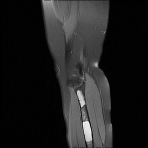
Diagnosis of “fibrous bone dysplasia” is possible even on the basis of the symptoms that appear, but instrumental examinations also play an important role, among which the main role is played by MRI. Therapy in all situations involves surgical intervention.
Pathogenesis
Until now, the basic prerequisites for development and the mechanism of the origin of pathology remain unclear to the end. However, according to most doctors, the source lies in gene mutations and malformations in the prenatal period.
So, experts believe that some predisposing conditions play an important role in the onset of the disease:
- hormonal disruptions;
- disorders of the formation and development of bones, as well as cartilage;
- abnormalities of the ligaments and muscles connecting the joints;
- genetic inheritance;
- unbalanced nutrition of a future mother - the lack of the necessary set of vitamins, minerals and nutrients in the diet;
- abuse of bad habits and harmful external factors that negatively affect the female body during the period of gestation;
- exacerbation of chronic pathologies or past serious infectious diseases during pregnancy;
- the use of certain drugs without prescribing a doctor in the early stages of bearing a baby;
- the effect of chemical or toxic elements on the body of a woman in position;
- too pronounced toxicosis;
- the presence of a future mother of serious gynecological pathologies associated with the uterus and appendages;
- water shortage.

As you can see, the basis of the risk group is newborn children, however, medicine knows situations where such an anomaly was first discovered in a person of mature age. It is noteworthy that in women this pathology is detected much more often than in the stronger sex.
Varieties
There are several forms of fibrotic dysplasia:
- monossal - a lesion of one bone, symptoms can occur at any age;
- polyossal - a pathological condition covers several bones, for example, a shoulder or hip, but only on one side of the body.
Today, the most common is the classification of Zatsepin, which provides for the distribution of defect into several main types.
- Intraosseous fibrotic dysplasia can occur in both polyossal and monoossal forms. Microscopic examination reveals focal lesions, but sometimes tissue can cover the entire bone. It is noteworthy that patients suffering from such a disease do not have strong bone changes.
- Total injury. This variety differs from intraosseous dysplasia in that all parts of the bone are affected, which causes deformation, and this, in turn, provokes regular fractures. The name of the pathology speaks for itself: it proceeds only in a polyossal form. Most often, total fibrous dysplasia of the femur and tibia occurs.
- Tumor type. This variety always provokes the proliferation of pathological tissue. Such dysplasia is extremely rare.
- Albright's disease. This type of pathology is considered the most common, most often diagnosed among children. The anomaly has a fleeting character and is rapidly progressing.
- Fibrocartilage variety. The peculiarity of this form is that almost always this pathology acquires malignant features.
- Calcifying type. A special kind of anomaly that is extremely rare.
Additional classifications
Pathology also differs in the localization of damage, most often abnormal changes include:
- jaw and skull;
- small and large tibia;
- humerus
- spine and ribs;
- knee-joint.
In addition, fibrous dysplasia of the skull has its own classification.
- The cystic form usually affects the lower jaw. Specific neoplasms are multiple and single.
- Sclerotic variety. It is characterized by the appearance of places with pronounced changes in bone tissue, because of which they are densified. Most often, such anomalies affect the base of the skull, frontal and maxillary bone, as well as the nose.
- Paget-like form. Pathological changes occur with Paget's syndrome and are based on an increase in the volume of the brain zone of the skull.
Clinical picture
The first foci of the disease arise in the early childhood and for a long time do not manifest themselves at all. However, with the growth of the child, the disease is constantly progressing. As a rule, after completion of the development of the child’s body during puberty, the course of the pathology becomes stable.
Only in exceptional cases, dysplasia continues to progress in adulthood. This phenomenon is due to the appearance of various complications or the presence of concomitant diseases.
The development of the pathological process provokes an increase in the volume of the injured area and squeezing nearby structures and organs, which leads to a violation of their functions. One of the initial symptoms of the defeat of the ENT systems can be a local inflammatory phenomenon, such as otitis media or sinusitis.
The first manifestations of the disease
Over time, the pathological lesion grows, increasing in size, while visible changes in the skeleton are formed with the presence of a compacted swelling. It is noteworthy that this site does not cause pain during palpation. The skin covering the tumor becomes thinned and too shiny due to its atrophy.
The patient may have complaints of general poor health, migraine, impaired vision or hearing. This applies to cases of development of skull dysplasia. If we are talking about other bones of the skeleton, then the patient most often notices pain and abnormal external changes.
Symptoms
Clinical signs of pathology differ significantly depending on the localization of the process itself. But some common features are found in all varieties of the disease. What do patients with fibrotic dysplasia look like? Photo of patients, to put it mildly. unpleasant, so we decided to refrain from publishing such images. If desired, the reader can always get acquainted with specialized literature or materials of purely medical portals. And yet, how to visually identify the signs of vice? Typically, changes in the patient's body are more than obvious.
For example, fibrous tibia dysplasia is accompanied by such symptoms:
- lameness when walking;
- regular fractures;
- shortening of the femur;
- breaks of the damaged area.
Dysplasia of the knee manifests itself differently:
- pain syndrome, the intensity of which increases significantly due to physical exertion or due to a sharp change in weather;
- partial or complete change in the shape of the legs;
- characteristic articular crunch during flexion or extension of the knee;
- change in the shape of the knee joint and calyx;
- increased knee mobility.
Pathology of the jaws stands out:
- facial deformity;
- thickening of the injured bone;
- slow puberty.
Fibrous dysplasia of the bones of the skull provokes the occurrence of such manifestations:
- tooth abnormalities;
- decrease in skull volume;
- the predominance of the brain area over the front part;
- curvature of the first vertebra;
- change in the paranasal sinuses.
Often this disease also leads to the spread of pathology to the hip joint and ridge, which provokes all kinds of disturbances in posture.
If an abnormality is found in the humerus, a disorder in the mobility of the injured limb occurs.
Albright's disease has the most pronounced symptoms:
- malfunctions of the endocrine apparatus;
- premature puberty in women;
- violation of the normal proportions of the body;
- focal skin pigmentation;
- complex bone changes.
In addition, this type of pathology is often accompanied by impaired functioning of internal systems and organs.
Diagnostics
A qualified person can easily make the correct diagnosis based on visual symptoms alone. Initial research may consist of:
- a detailed survey of the patient, necessary to determine the time of occurrence of the initial signs and the intensity of their severity;
- studying the history of pathology and the existing history - to search for a factor predisposing to the occurrence of a disease;
- careful physical examination of the injured area, its palpation and percussion.
The detection of dysplasia does not imply laboratory tests of urine, blood and feces, since they do not have diagnostic value in this case.
Among the instrumental methods for detecting the disease can be identified:
- MRI
- densitometry;
- CT
- radiography.
Differential diagnosis is very important, which will help to separate the pathology from all kinds of tumors, inflammatory and hyperplastic phenomena.
In addition, the diagnosis can be confirmed by endoscopic examination with a biopsy. In this embodiment, the taken tests are additionally sent for histological testing.
Although in reality, obvious changes in the pathological nature are clearly visible on x-rays. Fibrous dysplasia can be detected even on the basis of this type of study and a detailed examination of the patient. But mild forms of the disease are still desirable to confirm with the help of auxiliary methods.
Treatment of fibrous bone dysplasia
The use of conservative methods of therapy, as a rule, does not bring a positive effect. That is why usually patients with a confirmed diagnosis of "fibrous dysplasia" surgery are prescribed without fail. This elimination of pathology is recommended for both adult patients and children.
The treatment of fibrotic dysplasia is carried out by:
- resection of the injured area with further replacement of the bone graft;
- osteotomy
- lengthening of the damaged bone - such an operation is necessary only in cases where the limb is shortened.
Treatment for complications
Polyostotic fibrotic dysplasia can significantly limit the ability of surgeons and take away the patient’s chance of performing an operation. In this situation, treatment with other methods is possible:
- physiotherapeutic manipulations;
- the use of special orthopedic shoes and other devices that reduce the load on damaged joints and bones;
- Exercise therapy;
- therapeutic massage courses.
Prevention
Due to the fact that the exact mechanism of the origin of the defect is still unknown, in order to prevent such a disease, it is necessary to adhere to the general rules:
- lead a full, healthy lifestyle while carrying a child and regularly visit a gynecologist;
- comply with all safety rules when working with toxic, chemical and other hazardous substances;
- follow a proper, balanced diet;
- keep your own body in shape;
- to exclude the effect of increased physical exertion on the most frequent places of the occurrence of pathology;
- Systematically undergo a full medical examination.
Forecast
The outcome of fibrotic dysplasia is usually favorable. Only a severe polyossal form of anomaly is capable of provoking the appearance of serious mutilating changes. Complications as the formation of a malignant neoplasm occur in only 4% of all cases of the disease.