In the article, we will look at what an ulcer of the skin and mucous membranes that develops in different organs looks like. The presented photos will help to recognize the onset of the disease in order to begin treatment procedures as soon as possible. You will find out the causes of their occurrence on the integument of the body, the main symptoms by which they can be recognized, how ulcers are treated.
First, let's find out how this type of tissue damage differs from simple wounds and erosion. An ulcer is a deep, inflamed, long-healing defect on the surface of the skin or mucous membrane, the healing of which occurs with scarring, since tissue particles are irretrievably lost.
Ulcers can be the result of an infectious disease, mechanical damage to the integument, occur after chemical or radiation exposure. Frequent ulcers from varicose veins or other disorders of the blood supply to the organ, as well as the innervation of the place. If the ulcer does not heal within one month, then it is already a trophic ulcer. Most often, you can see what a trophic ulcer looks like on the lower limbs (feet and lower leg) of people suffering from impaired venous outflow. Irreversible processes on the skin lead to the fact that the protective function of this place is greatly weakened and is not able to withstand external influences.
Next, we will examine in more detail what the ulcer looks like in different places of the human body, how to recognize it at an early stage according to common symptoms, and how to treat it.
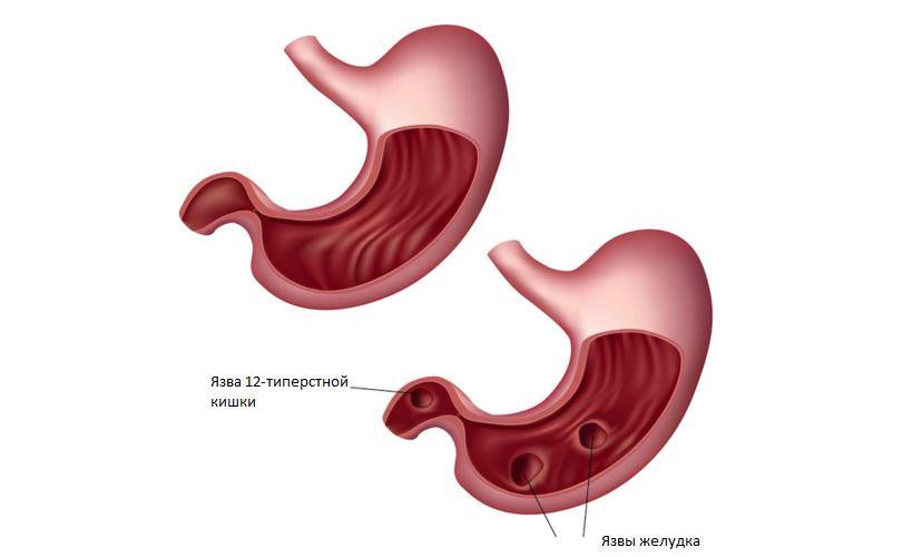
An ulcer on the walls of the stomach
The main cause of gastric ulcer is considered to be the effect of Helicobacter pylori. This is a bacterium that enters the stomach with food and multiplies rapidly, harming the mucous membrane. The ability of protective mechanisms is reduced, and the mucus secreted by the stomach is no longer able to cope with pepsin and hydrochloric acid. Aggressive factors destroy the mucous membrane, which leads to ulcers. Another reason for the appearance of an ulcer in the stomach is considered to be a nervous shock, constant stresses that cause muscle spasms and, of course, gastrointestinal blood vessels.
As a result of a violation of blood flow in the stomach, congestion is observed, the aggressive environment of the gastric juice corrodes the walls, which leads to an ulcer. Also, the disease can also occur as a result of other diseases - tuberculosis or diabetes, cirrhosis of the liver or hepatitis, lung cancer or pancreatitis, etc. Injuries to the stomach of a mechanical or chemical nature can also bring the mucosa to ulcers, like some medications.
Diagnosis of the disease
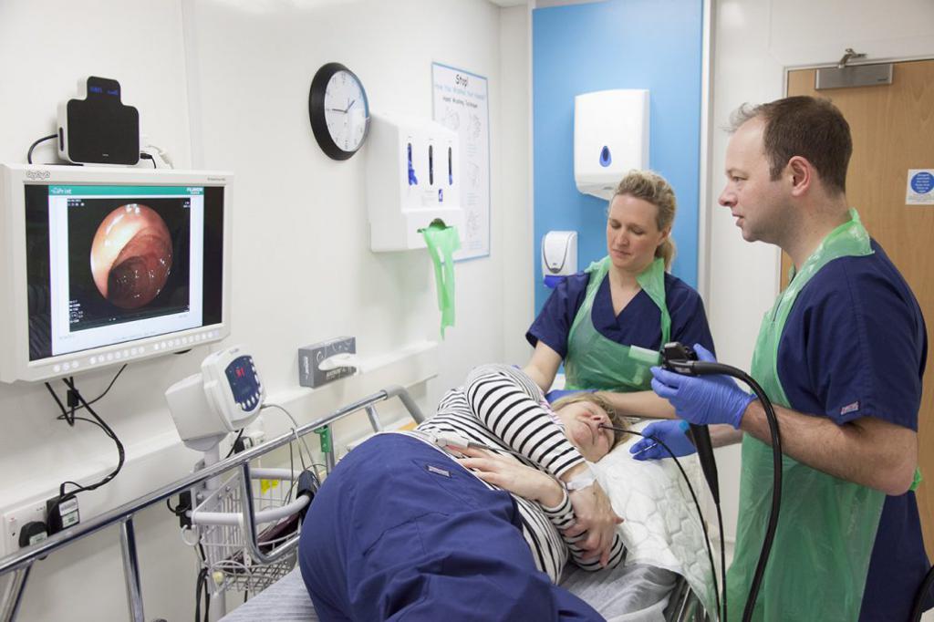
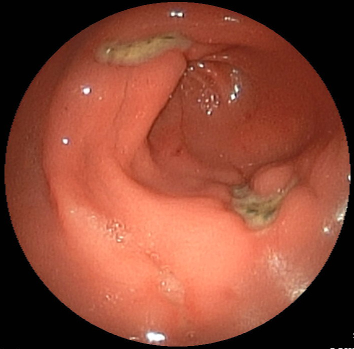
The doctor is able to discern what a stomach ulcer looks like by making the patient fibroesophagogastroduodenoscopy. This is a study during which a small chamber descends through a special tube into the stomach and the entire mucous membrane with all the flaws is visible on the monitor screen. At the same time, the contents of the stomach are taken for analysis, checking for the presence of Helicobacter pylori. Also, the doctor prescribes blood, urine and feces tests to detect blood impurities. Be sure to make the patient an ultrasound of the digestive tract, as well as radiography with contrast or monitoring the pH of the gastric juice, checking it for aggressiveness of the environment.
What a stomach ulcer looks like, the patient will not be able to see, but he will understand its presence in the body by the symptoms. These are painful sensations in the stomach, heartburn after eating, burning, nausea, excessive gas formation with belching, a feeling of fullness even after a small portion of food. With the disease, appetite decreases, there is a feeling of heaviness in the stomach, there is a violation of the stool (there can be both constipation and frustration).
How to cure a stomach ulcer
Look carefully at the photo of what a stomach ulcer looks like. Mucous can be damaged in one or in several places. If the ulcer bleeds or penetrates through the entire wall of the stomach, then surgery is prescribed. If the ulcer is not perforated, then the treatment consists of several stages.
- Antibiotic therapy. A doctor prescribes at least two antibiotics from the tetracycline or penicillin group.
- Medicines that are designed to enhance the protective properties of the mucosa, for example, De-Nol, which forms a film on the walls of the stomach.
- Antisecretory drugs not only envelop the mucous membrane, but also neutralize the effect of aggressiveness of hydrochloric acid. At the same time, heartburn, gas formation and pain are significantly reduced. It can be Maalox or Almagel.
- Proton pump blockers block the increased formation of the corroding stomach wall of hydrochloric acid. This is Omez or Omeprazole.
- In addition, the doctor may prescribe probiotics (Linex or Bifiform), a sedative effect after eating gives Valerian, improves the motor functions of the digestive organs Motilium, and relieves tension and spasms of No-shpa.
The treatment is carried out for a rather long period. At the same time, a strict diet is observed, excluding alcohol, coffee, broths, flour products, canned food, seasonings, smoked meats and fatty meat and fish, fried foods and vegetables that cause gas formation (beans, legumes, radishes and cabbage) are not allowed. Fruits and vegetables should be boiled. And citrus fruits should be generally excluded due to acid, which will negatively affect the mucous membrane.
Duodenal ulcers
What does a duodenal ulcer look like? Just like the stomach. Symptoms of the disease are also similar, as are the causes of the disease. Treatment is carried out for the recommendations of a gastroenterologist. A strict diet must be observed, as the following complications are possible:
- bleeding from an ulcer site;
- perforation, that is, an ulcer passes through the entire wall of the intestine;
- penetration, when the spread of the ulcer goes to nearby organs;
- pyloric stenosis, in which the opening between the stomach and intestines becomes narrow, which interferes with the passage of food from one organ to another.
To avoid ulcers of both the stomach and the duodenum, preventive measures must be taken. Lead a healthy lifestyle, eat right, avoid stressful situations, do not drink alcohol, do not smoke. Observe the regime of the day, refuse tedious work, do not go out on a night shift. It is imperative to follow a strict diet so that the ulcer does not develop into a chronic form.
White plaque in the mouth
If ulcers appear on the mucous membrane of the mouth or gums, this may indicate not only the appearance of a dental disease, but also many other infectious diseases, including syphilis and HIV. Many have seen mouth ulcers. In the photo below, you can consider it more closely.
Such ulcers have rounded outlines, inflamed borders and a white coating on the wound itself. Her presence in the mouth brings discomfort during eating, drinking, talking. A person experiences pain, increased salivation, general weakness, loss of appetite and even an increase in body temperature, the causes of which are:
- stomatitis;
- gingivitis;
- periadenitis in necrotic form;
- tuberculosis of the mucous membrane of the mouth or lungs;
- infections
- oncological diseases;
- herpes or enterovirus infection;
- injury during dental operations, from biting the cheek or blow to the jaw, chemical burns or reaction to too acidic food, from rough bristles on the toothbrush;
- as a result of exposure to drugs.
What the ulcer looks like can be seen in the mirror. If at home it is not possible to cure such an ulcer, then it is necessary to consult a specialist and identify the root cause of the disease. For treatment, drugs are used based on the results of the examination. It can be antifungal or anti-inflammatory, antihistamines or antiviral drugs. If a herpes virus is found, then anti-herpes substances are prescribed. A complex of vitamins and minerals will also help. A good help in the complex treatment will be folk methods - decoctions of herbs: chamomile, sage or eucalyptus. Local anesthetics in the form of ointments and sprays will reduce pain. After treatment, the doctor needs to monitor what the ulcer looks like, whether there are any improvements in a positive direction.
Preventive actions
To minimize cases of stomatitis and other ulcers in the oral cavity, you need to follow the rules of hygiene - use a high-quality toothbrush, monitor the health of the oral cavity. It will help strengthen the body's immunity in general, eating fruits and vegetables. Nutrition should be balanced, containing all the vitamins and minerals necessary for the body.
Irrigators are considered an effective method of cleaning the oral cavity from food debris. Under the strong pressure of water, you can free all the gaps between the teeth, preventing both stomatitis and periodontal disease of the gums.
Skin ulcers
The human epidermis tends to recover quickly from damage. But there are cases when the rehabilitation processes are significantly slowed down or completely stopped. Places in which necrotic tissues fall away, but new ones do not grow, turn into non-healing skin ulcers. How this defeat looks and develops in stages, can be seen in the figure below.
Tissues in such places do not recover for a long time, skin ulcers cause a lot of inconvenience. Slow skin regeneration depends on the weakening of the human immune system, the presence of inflammatory processes. Yes, and the ulcers themselves become the entrance "gate" for many bacteria. Infections quickly enter the circulatory system and spread throughout the body.
Causes of occurrence
- Various injuries associated with both mechanical damage to the integrity of the skin, and with electrical, thermal or radiation exposure.
- Ulcers on the skin can occur from tumors, for example, sarcoma or lymphogranulomatosis.
- With blood flow disorders, anemia, blood diseases, as well as scurvy or diabetes mellitus, side effects in the form of skin ulcers are possible. What leg ulcers look like can be seen in a patient with varicose veins.
- Infectious skin lesions.
- Progressive paralysis.
- Diseases associated with changes in the structure of the walls of blood vessels, for example, Raynaud's disease or syphilitic aortitis.
External ulcer treatment
Having examined how the ulcers look in the photo, the patient can go to the doctor in time and begin treatment. The first step is a series of hygiene measures. The surface of the skin is cleansed of purulent discharge, formulations or ointments stretching purulent contents are applied to the wound. Dressings need to be changed several times a day.
Well affect folk skin methods:
- You can wash the ulcer using freshly squeezed juice from leaves of cabbage or potatoes.
- Fresh wormwood, ground into fresh pulp, is applied to a bandage folded several times, and applied to hardly healing ulcers, changing dressings 2 to 3 times a day.
- Juice of room geranium or extract of comfrey also heals affected areas of the skin well.
In combination with hygienic and cleansing procedures, you need to increase immunity. For this, vitamin preparations are prescribed.
If external treatments do not help get rid of the ulcer, then surgery is recommended. During the operation, dead skin is cut off, a deep defect is filled with a graft.
What do ulcers look like with varicose veins?
Varicose veins are accompanied by the destruction of the structure of blood vessels. From this, an insufficient amount of nutrients and oxygen enter the epidermis. To determine how the ulcers on the legs look, you can visually examine the limbs. If a constantly wetting wound appears, you should definitely consult a doctor, because varicose ulcers often develop into trophic ones. They can occur not only from varicose veins, but also with diabetes.
The skin in the affected area acquires a burgundy shade, becomes inflamed, begins to itch. You can see heavily pigmented spots, bruises, palpation felt compaction on the skin. These are all the precursors of the appearance of varicose ulcers.
If you have these signs and see what the leg ulcer looks like in the photo, immediately rush to the doctor to start treatment on time, because these ulcers have a number of complications, such as damage to not only all layers of the skin, but also muscle tissue, tendons and even bones .
Varicose ulcer treatment
First of all, treatment begins with the treatment of the ulcer with antiseptics. This will inhibit the suppuration and growth of bacteria. They treat wounds several times a day with Furacilin or Miromistin.
To skin regenerated faster, you need to use "Lemikol". It will not only calm the inflammatory process on the skin, but also relieve irritation on the surface. This ointment effectively heals wounds.
Anti-inflammatory and bactericidal drugs, general-purpose vitamin complexes will help accelerate recovery. It is important to strengthen the body's immunity so that it is actively involved in the fight against the harmful effects of microbes.
Venotonics are simply indispensable in the treatment of varicose veins. They contribute to the regeneration of blood vessels, exclude the formation of blood clots.
What does a trophic ulcer look like? Usually it is suppurative. The effect of Argosulfan is already needed here. This is a strong antibacterial drug that is used externally to fight ulcers.
Often, ulcers with varicose veins constantly itch, causing an irresistible desire to comb a sore limb. This can be detrimental. To reduce itching, you will need antihistamines such as Suprastin, Fenistil, etc.
Physiotherapeutic procedures are also used in the treatment - laser or ultraviolet radiation. During the session, the wound is dried and microorganisms die.
How to identify a trophic ulcer
Most trophic ulcers are associated with impaired blood flow. Most often, they outgrow from venous ulcers and are localized either on the foot or in the lower parts of the lower leg. If a trophic ulcer appears on the leg (what the clinical picture looks like, we will describe below), you should immediately consult a doctor for help, without bringing the matter to irreparable consequences or surgical intervention.
The formation of a trophic ulcer proceeds gradually. The skin on the leg becomes crimson and darkens, you can feel the seals and swelling. At the site of the future wound, the epithelium stretches and becomes brilliant. Specialists gave the process the name "patent leather." Drops of lymphatic fluid appear on it. Even minor skin damage can lead to wound formation.
Gradually, its size increases both in breadth and in depth. The process is accompanied by intolerable itching, pain, often an increase in body temperature. An infection gets into the wound and suppuration begins. Sometimes the whole body suffers from a trophic ulcer, a general infection is obtained, which can cause fatal consequences for a person.
Treatment
What a trophic ulcer looks like in the photo, you already know. Consider methods of treating such a skin pathology. First of all, the patient eliminates the cause of venous insufficiency. First of all, it is necessary to stop the increased pressure and edema at the site of the ulcer, as well as the pathological return of blood flow through the veins.
Laser and radiofrequency thermoagulation are considered modern and quite effective ways to establish the correct venous outflow, remove sclerotization of damaged vessels.
After this, the doctor examines what the trophic ulcer on the leg looks like (photo at the beginning of the article) and prescribes venotonics and medications that enhance skin regeneration in this place. If the ulcer has a diameter of more than 6 cm, then transplantation of material from the thigh or from the front wall of the abdomen is practiced on the skin. In some cases, a fasciotomy is performed. Dissection of muscle tissue allows you to ease the pressure inside the tissues, while blood circulation is facilitated.
Anti-inflammatory drugs and physiotherapy will help. Treatment with polarized light will help relieve pain, reduce itching, and accelerate recovery. After eliminating the acute condition, the patient will be recommended elastic bandaging of the limb or wearing compression underwear. This will relieve stress and improve the overall condition of the legs. In order for the bloodstream to better disperse blood through the vessels, the doctor will advise you to take daily walks and increase physical activity. If you are tired, you need to put your legs on a raised platform, for example, lying on a sofa, raise them on a pillow.
anthrax
This is a very dangerous infectious disease that is transmitted to humans from animals, both agricultural and wild. It is characterized by fulminant course and hemorrhagic inflammation on the skin or internal organs, a carbuncle with purulent contents appears. Infection of anthrax can occur after contact with a sick animal, through flies of a zhigatka or bites of horseflies, less often it can be infected through soil or food. How does anthrax look like, should know farm workers and slaughterhouses, skin makers at enterprises.
The clinical picture is characterized by acute manifestations - high fever, severe chills, swollen lymph nodes. At the point of entry of the infection, a stain is formed first, similar to an insect bite. It hurts a lot, acquires a red-blue color, constantly itches. A day later, the stain passes into the papule, then into the vesicle a few millimeters in diameter, which is filled first with serous and then bloody fluid. When an inflated bubble bursts, an ulcer remains on the skin. Small vesicles appear on its external contours, which expand the ulcer. This process lasts 2-3 weeks. A black scab (necrosis) forms in the center of the ulcer. Swelling and a granulating ulcer appear. Fatty tissue is captured under the skin and the process ends with scab rejection and the formation of a dense scar on the skin.
For treatment, in addition to the antibiotics of the penicillin group, an antisybrasyl immunoglobulin is prescribed. Treatment should only take place in a hospital and under strict medical supervision. In order to prevent such a terrible infection, special measures are applied on farms and on large farms - they reorganize and vaccinate animals on time, observe hygiene standards. If nevertheless there was contact with a sick animal, it is necessary to undergo prophylactic antibacterial therapy for 5 days.
The article examined in detail all types of ulcers, the photo shows how they look on the skin and on the mucous membrane of the stomach and oral cavity. Take care of yourself and do not pull with a trip to the doctor. After all, it is much easier to recover in the early days of the disease than from its neglected form.