In the kidneys, as in any other organ, the formation of various cysts is possible. Of these, only about 4% of diagnosed cases are congenital pathologies, the rest are formed in the process of growing up a person. One of the varieties of such neoplasms is a subcapsular cyst of the kidney. Hearing this diagnosis, many patients begin to panic and despair, but whether it is as dangerous as they are imagined is worth sorting out.
What is a subcapsular kidney cyst?
In the photo, a subcapsular kidney cyst, which is presented in a schematic image. A neoplasm may consist of one or two chambers. Often its size does not exceed more than 2 cm, but with rare exceptions, the cyst grows to 10 cm.
The surface layer of the kidney is a fibrous tissue that has the ability to stretch. It creates a certain protective capsule, in which the organ itself is located. Under the influence of provoking factors, a neoplasm is formed between the outer membrane and the parenchyma of the kidney, which is subsequently filled with fluid and is called a subcapsular cyst. Its shape may resemble a ball or an ellipse. If the cause of the growth was trauma, then the internal fluid will contain impurities of pus and blood.
The danger of the disease is that at the initial stage it is completely asymptomatic, as small subcapsular kidney cysts do not affect blood vessels, pelvis, and also do not interfere with the formation and excretion of urine. With a combination of provoking factors, there is a likelihood of a benign cyst becoming malignant. Therefore, this disease belongs to the category of potentially dangerous pathologies for human health.
Symptoms of the pathological process
It is not always possible to detect the formation of a subcapsular cyst of the left kidney, as well as the right one. Often this process is completely asymptomatic.
But in some cases, the following symptoms are possible:
- aching dull pain that appears as a result of squeezing the kidney with adjacent tissues against the background of an increase in its size during the formation of a cyst;
- a feeling of heaviness in the right or left side, depending on the location of the cyst, which is associated with a heavier organ due to the large volume of fluid in the neoplasm;
- elevated blood pressure, as an enzyme, rhinin, is released during the formation of a cyst;
- frequent pathologies of the urinary system of an infectious property;
- failure of the outflow of urine;
- compaction in the abdominal cavity;
- an increase in kidney size, which can be detected by palpation;
- in the urine, there are impurities of blood against the background of increased intrarenal pressure.
If at least one of the symptoms appears, you must consult a doctor and undergo a full examination. This will help refute suspicions or identify a pathological process before complications appear, which will make it possible to affect the growth of the cyst without surgery.
Reasons for education
The main reason for the formation of a subcapsular kidney cyst is the excessive growth of the epithelium inside the canal. It can provoke a metabolic failure in the body. As a result of this, exfoliated cells of the inner layer clog the canal and prevent the natural outflow of urine. As a result of this, it increases, ceases to participate in the work of the kidney, a cyst forms.
Other causes of subcapsular kidney cysts:
- necrosis of a separate section of the organ, while the often formed cyst resolves independently;
- congenital anomalies as a result of impaired fetal development in the womb;
- injuries
- complication of diseases (pyelonephritis, glomerulonephritis).
Any change in the structure of the organ leads to a failure of its functionality. This can subsequently provoke the formation of a subcapsular cyst of the right kidney, as well as the left.
Diagnostics
To establish a diagnosis, the doctor prescribes various studies that help determine the location of the cyst, its size and stage of development. Laboratory tests are also used in the collection of history. But they can only determine the general condition of the paired organ, the degree of its functionality and possible violations, however, they are not able to identify the cyst.
The following types of studies are more informative:
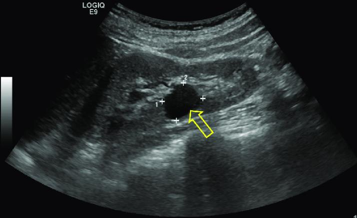
- Ultrasound The study is carried out in order to identify neoplasms in the organ, determine their quantity, size. Ultrasound of the subcapsular kidney cyst helps to identify the pathological process in the fetus already at the 15th week of pregnancy.
- MRI The examination is auxiliary when there are certain inaccuracies after ultrasound. Using it, you can identify even the most minor changes in the structure of the kidney, determine their nature and nature of occurrence.
- Antiography is one of the varieties of computed tomography using a contrast agent. With the help of this study, not only the structure of the kidney is determined, but also the state of adjacent tissues, vessels and arteries is studied. The greatest contrast appears in the most important areas of the organ, which allows you to see the defects. The catalyst is often iodine.
- Radioisotope study. This study helps to identify a cyst at an early stage of development, when ultrasound could not detect it. To carry it out, a small amount of isotopes is introduced into the body, which are rapidly eliminated. Their concentration depends on the weight, age and height of the patient. The level of exposure in a radioisotope study is much less than with ultrasound.
The subcapsular cyst of the kidney, both of the right and the left organ, does not have any differences, but when the diagnosis is made, the doctor will definitely clarify the true localization of the growth, which is necessary for further treatment.
Drug treatment
If the diagnosis showed the benign nature of the cyst, its small size and simple structure, then medications are prescribed for treatment. Most often, their action is aimed at eliminating unpleasant symptoms and facilitating the well-being of the patient.
For these purposes, the following types of drugs are used;
- painkillers;
- antimicrobial agents;
- antibiotics
- drugs to lower blood pressure;
- drugs that reduce the concentration of calcifications in the kidneys.
More accurate information about the regimen and dosage of drugs indicates the attending physician based on the established diagnosis.
Congenital pathological process requires control over the water balance in the body. Therefore, a person should drink at least 2 liters of water daily and take medications to lower blood pressure.
Drug therapy is acceptable only if the cyst is benign, so it is forbidden to experiment and use drugs on your own without the knowledge of a doctor.
Ambulatory therapy
This method of treating a subcapsular kidney cyst is used to detect a larger benign neoplasm. In this case, the doctor decides to conduct a less traumatic surgery to remove the growth.
The main ways:
- Drainage or puncture. An indication for carrying out is the presence of a growth of more than 6 cm. During surgery, a thin tube is inserted into the neoplasm to drain the fluid contained in it. And the resulting emptiness is filled with a special solution that glues the walls of the cyst. Often, alcohol is used for this. After surgery, additional inpatient treatment is not required for the patient.
- Retrograde intrarenal removal. During the procedure, an endoscope is inserted into the urethra, which subsequently passes through the bladder and ureter into the affected kidney. Then, the affected area is dissected with a laser and a cyst is removed. After this, the wound is sutured.
Surgery
In some cases, surgical removal of the cyst cannot be avoided. The probability of the operation is determined by the doctor based on the possible risks to the patient's life.
The main indications for conducting:
- high blood pressure that cannot be stabilized with drugs;
- degeneration of the growth into a malignant formation;
- acute pain that cannot be eliminated;
- critical dysfunction of the affected kidney;
- rapid cyst growth.
To completely remove the cyst, laparoscopy is performed. During the operation, 2 small incisions are made: on the anterior abdominal wall and on the side of the affected kidney. One hole is necessary for introducing a camera with a lighting device, and the second for a removal tool. The number of incisions may be more, at the discretion of the surgeon.
At the end of the procedure, the patient stays in the hospital for another 3-5 days to monitor the dynamics of his health.
Folk remedies
On the network you can find recommendations on the use of folk remedies for the treatment of subcapsular kidney cysts, but they are not able to help the situation.
No herbs and decoctions can eliminate the neoplasm. And their use will only delay the process and miss the time for treatment, which will lead to the degeneration of a benign tumor into a malignant one.
Possible complications
The greatest danger is the ability of degeneration of the cyst into a malignant neoplasm. According to statistics, this occurs in 30% of diagnosed cases.
In addition, the following types of complications of pathology are possible:
- blood poisoning (peritonitis);
- internal bleeding;
- acute intoxication of the body;
- violation of the functionality of the body.
The faster the cyst is detected, the less harm it will cause to the body. Therefore, for any alarming symptoms, you should consult a doctor.
Forecast
Congenital pathology has an unfavorable prognosis. Most often, the child's life expectancy is no more than 2 months.
The acquired form of the cyst is treated, and the prognosis is favorable regardless of the method of treatment. Provided that the diagnosis will be carried out in a timely manner.
Prevention
There are some rules that can help avoid a recurrence, and also reduce the likelihood of the initial appearance of a subcapsular kidney cyst.
Key recommendations:
- timely treat kidney disease, preventing them from becoming chronic;
- avoid hypothermia and injuries;
- give up excessive drinking;
- minimize the consumption of harmful products;
- balance the diet by enriching it with fresh vegetables and fruits.
A careful attitude to your health will help to identify the pathological process at the initial stage. This will help to conduct timely treatment and prevent the transformation of the cyst into a malignant tumor. Therefore, even with minimal suspicion, it is recommended not to waste time, but to undergo an examination.